. 2022 Aug;92(2):279-291.
doi: 10.1002/ana.26380. Epub 2022 May 25.

ZSCAN1 Autoantibodies Are Associated with Pediatric Paraneoplastic ROHHAD

Affiliations

ZSCAN1 Autoantibodies Are Associated with Pediatric Paraneoplastic ROHHAD

Caleigh Mandel-Brehm et al. Ann Neurol. 2022 Aug.

Abstract

Objective: Rapid-onset Obesity with Hypothalamic Dysfunction, Hypoventilation and Autonomic Dysregulation (ROHHAD), is a severe pediatric disorder of uncertain etiology resulting in hypothalamic dysfunction and frequent sudden death. Frequent co-occurrence of neuroblastic tumors have fueled suspicion of an autoimmune paraneoplastic neurological syndrome (PNS); however, specific anti-neural autoantibodies, a hallmark of PNS, have not been identified. Our objective is to determine if an autoimmune paraneoplastic etiology underlies ROHHAD.

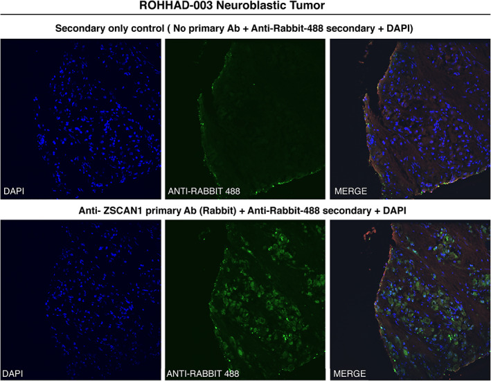
Methods: Immunoglobulin G (IgG) from pediatric ROHHAD patients (n = 9), non-inflammatory individuals (n = 100) and relevant pediatric controls (n = 25) was screened using a programmable phage display of the human peptidome (PhIP-Seq). Putative ROHHAD-specific autoantibodies were orthogonally validated using radioactive ligand binding and cell-based assays. Expression of autoantibody targets in ROHHAD tumor and healthy brain tissue was assessed with immunohistochemistry and mass spectrometry, respectively.

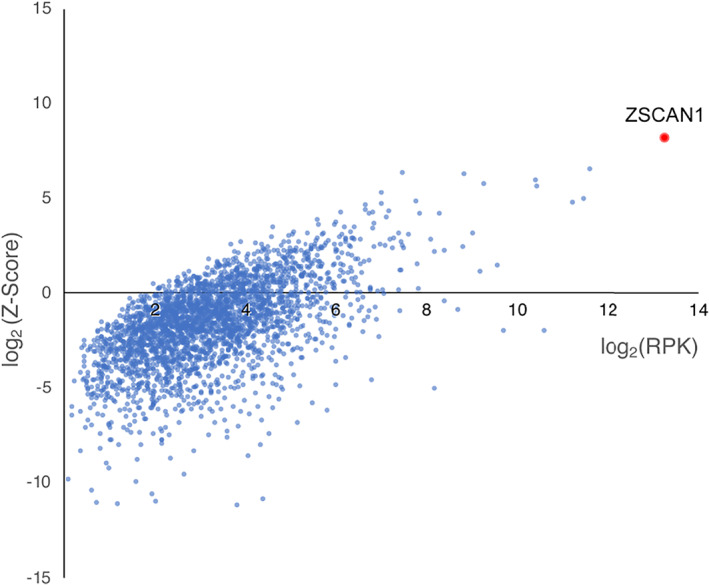
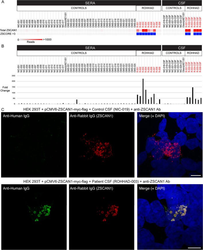
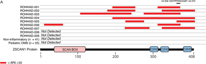
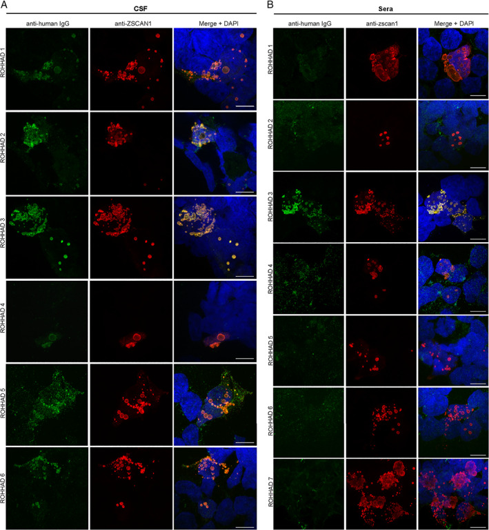
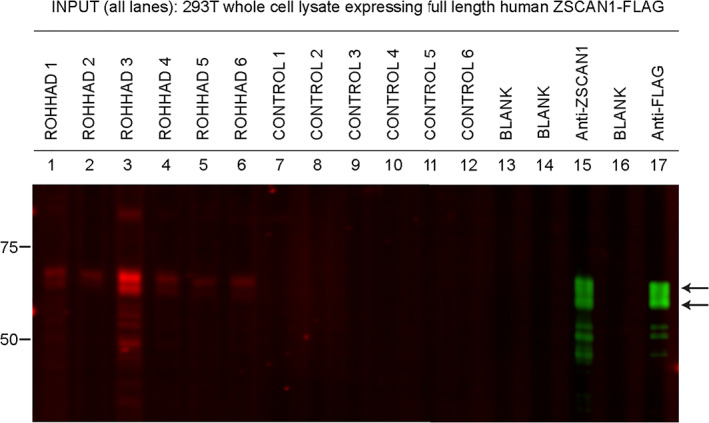
Results: Autoantibodies to ZSCAN1 were detected in ROHHAD patients by PhIP-Seq and orthogonally validated in 7/9 ROHHAD patients and 0/125 controls using radioactive ligand binding and cell-based assays. Expression of ZSCAN1 in ROHHAD tumor and healthy human brain tissue was confirmed.

Interpretation: Our results support the notion that tumor-associated ROHHAD syndrome is a pediatric PNS, potentially initiated by an immune response to peripheral neuroblastic tumor. ZSCAN1 autoantibodies may aid in earlier, accurate diagnosis of ROHHAD syndrome, thus providing a means toward early detection and treatment. This work warrants follow-up studies to test sensitivity and specificity of a novel diagnostic test. Last, given the absence of the ZSCAN1 gene in rodents, our study highlights the value of human-based approaches for detecting novel PNS subtypes. ANN NEUROL 2022;92:279-291.


Conflict of interest statement

Nothing to report.

Figures

FIGURE 1
FIGURE 1
PhIP‐Seq screen implicates ZSCAN1 as a candidate autoantigen in ROHHAD. CSF from 3 ROHHAD patients (ROHHAD‐1 to ‐3) and plasma from a large set of “healthy controls” (n = 100) were tested by PhIP‐Seq. Individual data were averaged according to cohort. All proteins with a ROHHAD mean RP100K >0 are plotted against ROHHAD Z‐score enrichments compared to healthy controls. Non‐ZSCAN1 proteins are denoted with blue dots; ZSCAN1 is denoted with a red dot.
FIGURE 2
FIGURE 2
Validation of autoantibodies to ZSCAN1 in ROHHAD patients. In panel a and b, enrichment of ZSCAN1 by PhIP‐Seq and RLBA was compared between ROHHAD patients (n = 9), non‐inflammatory healthy controls (n = 24), and pediatric controls including children with OMS with and without NT (n = 25) and an obesity patient with NT (n = 1). Data represent the average of 2 independent technical replicates. a, PhIP‐Seq analysis. Each column represents an individual sample. A heatmap of total ZSCAN1 RP100K is shown in the top row. To enable comparisons between the majority of samples with lower signal we added a ceiling value of 1,000 RP100K. Z‐Score enrichments based on our 100 human donor dataset are plotted below, with samples that have Z‐score enrichments >3 colored with blue squares. Gray squares indicate Z‐score <3. b, RLBA testing immunoprecipitation of recombinant ZSCAN1 by ROHHAD patients and controls. For all samples, fold change was calculated by dividing by the mean value from control sera (n = 17, mean = 20.83). c, Representative image showing immunostaining of 293T cells expressing full‐length ZSCAN1 with human CSF and commercial antibody to ZSCAN1 (rabbit). Top row shows immunostaining with control CSF. Bottom row shows staining with ROHHAD‐3 CSF. Colocalization is indicated by yellow in the merge images (far right both rows).
FIGURE 3
FIGURE 3
Peptide‐level ZSCAN1 enrichments by ROHHAD patients, informed by PhIP‐Seq. Cartoon graphic of the 408 amino acid ZSCAN1 protein with annotated SCAN and C2H2 domains is depicted below. Horizontal tracks above ZSCAN1 represent peptide enrichment data from individual ROHHAD patients or aggregated data from control cohorts. All peptides belonging to ZSCAN1 with enrichment RP100K >50 were plotted as red bars, merging together peptides with overlapping regions within individual tracks to reflect span of antigenic area. The black bar above all tracks represents an 11 AA region of overlap in 100% (7/7) patients within the C‐terminal domain. The amino acid sequence for the region of overlap is depicted above the black bar.
FIGURE 4
FIGURE 4
Validation of ZSCAN1 autoantibodies in CSF and sera of ROHHAD patients using 293 T CBAs. (A) CSF: Immunocytochemistry with 293T cells expressing full‐length ZSCAN1 and immunostaining with CSF (1:10) from ROHHAD patients and commercial antibody to ZSCAN1 (rabbit, 1:1000 Invitrogen). Anti‐human IgG‐488 was used to visualize human IgG, and anti‐rabbit IgG‐567 was used to visualize anti‐ZSCAN1 commercial antibody. Exposure times and post‐image processing and thresholding was kept constant across conditions within the experiment. Co‐localization was assessed qualitatively through observance of yellow in merged images. (B) Sera: Immunocytochemistry with 293T cells expressing full‐length ZSCAN1 and immunostaining with sera (1:100) from ROHHAD patients and commercial antibody to ZSCAN1 (rabbit, 1:1000 Invitrogen). Anti‐human IgG‐488 was used to visualize human IgG, and anti‐rabbit IgG‐567 was used to visualize anti‐ZSCAN1 commercial antibody. Exposure times and post‐image processing and thresholding was kept constant across conditions within the experiment. Co‐localization was assessed qualitatively through observance of yellow in merged images. Note colocalization in ROHHAD Sera‐1 and ‐3.
FIGURE 5
FIGURE 5
Detection of ZSCAN1 autoantibodies with slot‐blot Western blotting using ROHHAD CSF. Whole cell‐lysates from HEK293T cells expressing transfected with full‐length human ZSCAN1 cDNA were separated on a 1‐well 4–12% Tris–HCl protein gel, transferred to a PVDF membrane and immunoblotted in a slot‐blot device (BioRad). Primary and secondary antibodies were added to lanes as indicated. Primary antibodies were loaded in lanes 1–12. Lanes 13 and 15 served as secondary‐only controls. Primary antibodies are as follows: lanes 1 through 7: ROHHAD 1 through 7 CSF (1:200); lanes 8 through 12: controls 1–5 CSF (1:200); lane 13: blank; lane 14: commercial antibody to ZSCAN1 (Sigma, rabbit, 1:2000); lane 15: blank, lane 16: commercial antibody to Flag (CST, rabbit, 1:2000). Secondary antibodies to visualize human IgG loaded in lanes 1–12: goat anti‐human IgG (LICOR680). Secondary antibodies to visualize commercial antibodies to ZSCAN1 and FLAG lanes 14 and 16: goat anti‐rabbit IgG (LICOR800).
FIGURE 6
FIGURE 6
Immunohistochemical detection of ZSCAN1 in NT associated with ROHHAD patient‐3. Fixed neuroblastoma tissue was immunostained with either (top) anti‐rabbit‐488 secondary alone or (bottom) primary antibody to ZSCAN1 (rabbit) and anti‐rabbit‐488 secondary. Green: anti‐rabbit‐488 secondary; blue: DAPI (4’‐6'diamidino‐2‐phenylindole) to identify nuclei.

References

    1. Katz ES, McGrath S, Marcus CL. Late‐onset central hypoventilation with hypothalamic dysfunction: a distinct clinical syndrome. Pediatr Pulmonol 2000;29:62–68. 10.1002/(sici)1099-0496(200001)29:1<62::aid-ppul10>3.0.co;2-m. - DOI - PubMed
    1. Ize‐Ludlow D, Gray JA, Sperling MA, et al. Rapid‐onset obesity with hypothalamic dysfunction, hypoventilation, and autonomic dysregulation presenting in childhood. Pediatrics 2007;120:e179–e188. 10.1542/peds.2006-3324. - DOI - PubMed
    1. Ibanez‐Mico S, Marcos Oltra AM, de Murcia LS, et al. Rapid‐onset obesity with hypothalamic dysregulation, hypoventilation, and autonomic dysregulation (ROHHAD syndrome): a case report and literature review. Neurologia 2017;32:616–622. 10.1016/j.nrl.2016.04.008. - DOI - PubMed
    1. Patwari PP, Wolfe LF. Rapid‐onset obesity with hypothalamic dysfunction, hypoventilation, and autonomic dysregulation: review and update. Curr Opin Pediatr 2014;26:487–492. 10.1097/MOP.0000000000000118. - DOI - PubMed
    1. Lee JM, Shin J, Kim S, et al. Rapid‐onset obesity with hypoventilation, hypothalamic, autonomic dysregulation, and neuroendocrine tumors (ROHHADNET) syndrome: a systematic review. Biomed Res Int 2018;2018:1250721–1250717. 10.1155/2018/1250721. - DOI - PMC - PubMed

Publication types