

Plasma Amyloid and in vivo Brain Amyloid in Late Middle-Aged Hispanics
- PMID: 35466933
- PMCID: PMC10361456
- DOI: 10.3233/JAD-210391
Plasma Amyloid and in vivo Brain Amyloid in Late Middle-Aged Hispanics
Abstract
Background: Determining amyloid positivity is possible with cerebrospinal fluid and brain imaging of amyloid, but these methods are invasive and expensive.
Objective: To relate plasma amyloid-β (Aβ), measured using Single-molecule array (Simoatrademark) assays, to in vivo brain Aβ, measured using positron emission tomography (PET), examine the accuracy of plasma Aβ to predict brain Aβ positivity, and the relation of APOE ɛ4 with plasma Aβ.
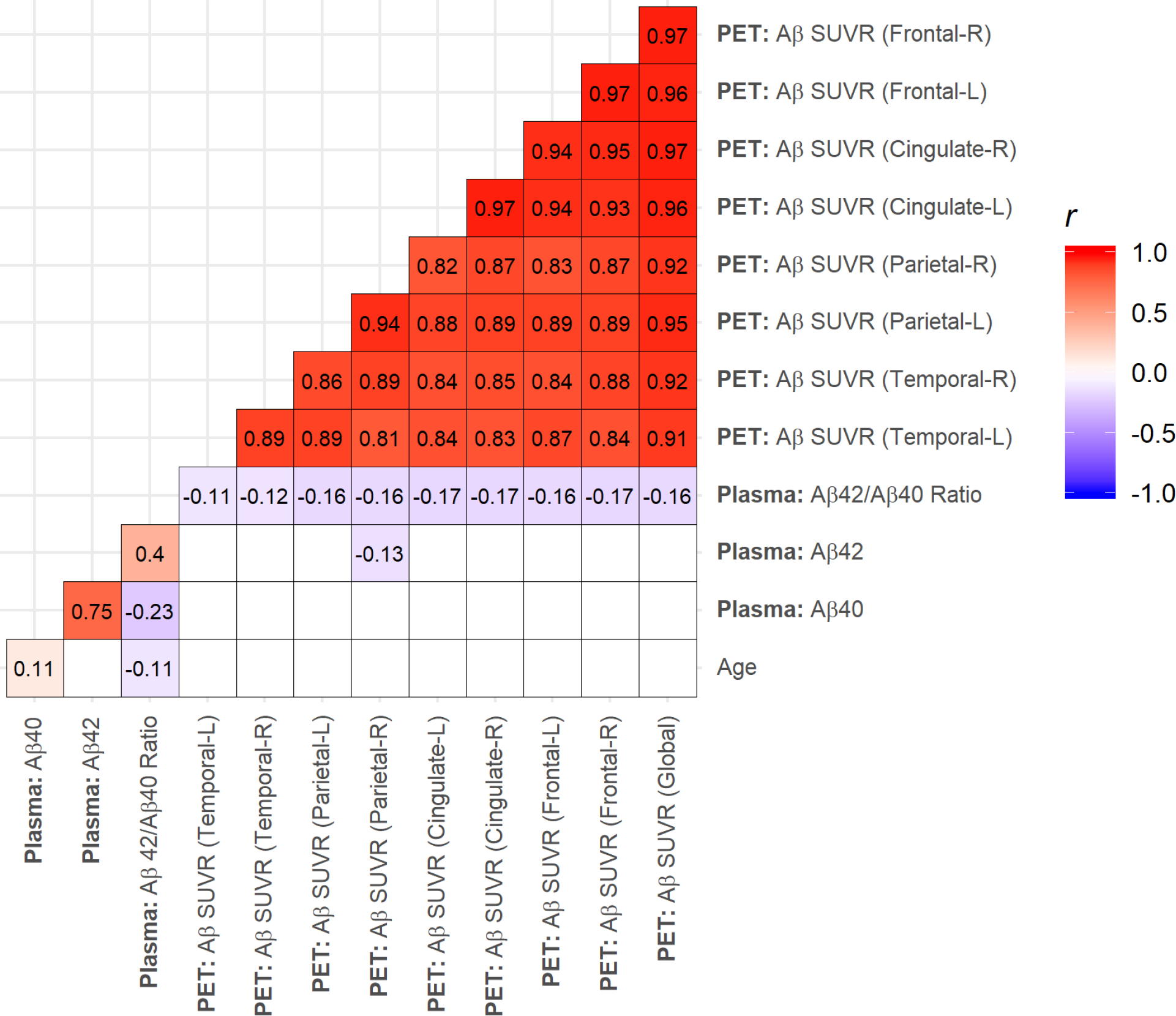
Methods: We performed a cross-sectional analysis in a cohort of 345 late middle-aged Hispanic men and women (age 64 years, 72% women). Our primary plasma variable was Aβ42/Aβ40 ratio measured with Simoa. Brain Aβ burden was measured as global SUVR with 18F-Florbetaben PET examined continuously and categorically.
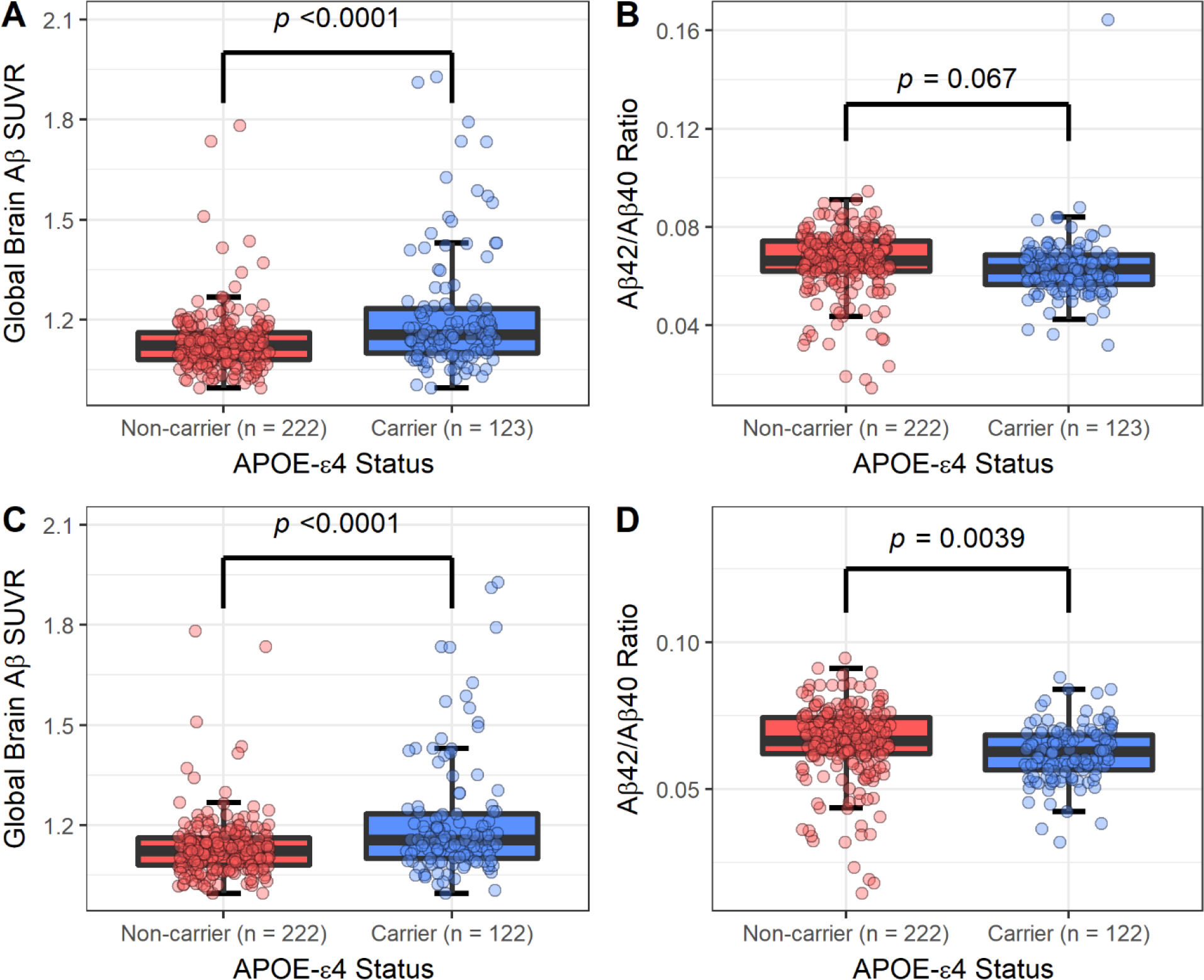
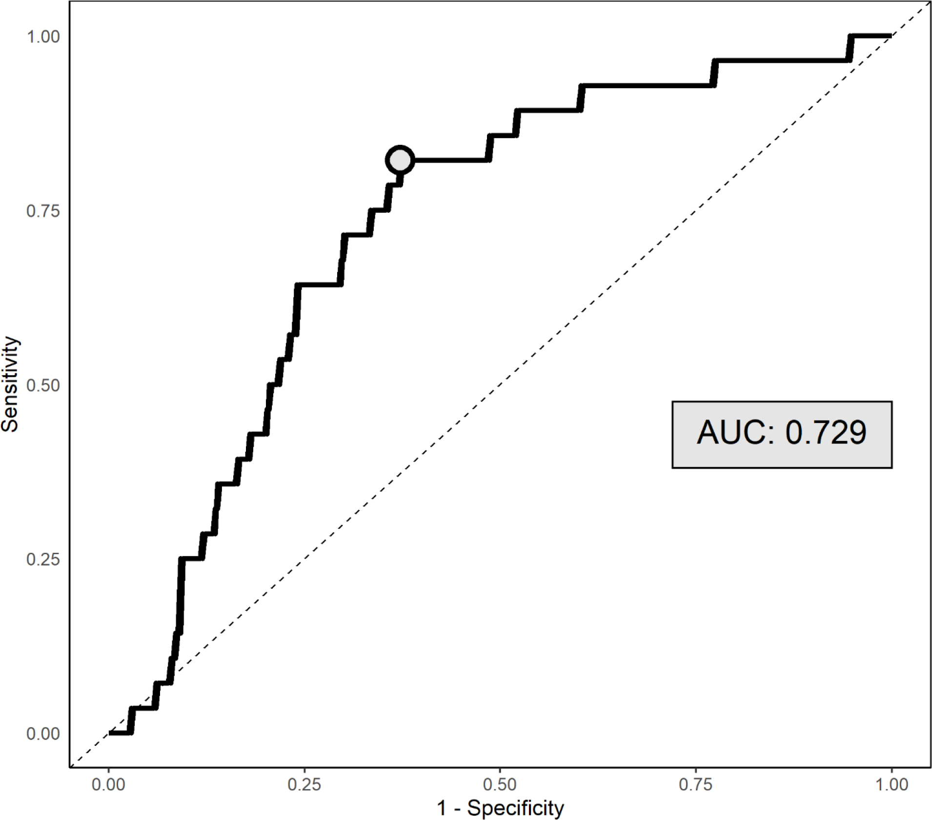
Results: Plasma Aβ42/Aβ40 ratio was inversely associated with global Aβ SUVR (β= -0.13, 95% Confidence Interval (CI): -0.23, -0.03; p = 0.013) and Aβ positivity (Odds Ratio: 0.59, 95% CI: 0.38, 0.91; p = 0.016), independent of demographics and APOE ɛ4. ROC curves (AUC = 0.73, 95% CI: 0.64, 0.82; p < 0.0001) showed that the optimal threshold for plasma Aβ42/Aβ40 ratio in relation to brain Aβ positivity was 0.060 with a sensitivity of 82.4% and specificity of 62.8%. APOE ɛ4 carriers had lower Aβ42/Aβ40 ratio and a higher Aβ positivity determined with the Aβ42/Aβ40 ratio threshold of 0.060.
Conclusion: Plasma Aβ42/Aβ40 ratio assayed using Simoa is weakly correlated with in vivo brain amyloid and has limited accuracy in screening for amyloid positivity and for studying risk factors of brain amyloid burden when in vivo imaging is not feasible.
Keywords: Alzheimer’s disease; amyloid; brain; hispanics; middle age; plasma; positron emission tomography.
Conflict of interest statement
CONFLICTS OF INTEREST
JA Luchsinger receives a stipend from Wolters Kluwer, N.V. as Editor in Chief of the journal
Figures
References
-
- Jack CR Jr., Knopman DS, Jagust WJ, Petersen RC, Weiner MW, Aisen PS, Shaw LM, Vemuri P, Wiste HJ, Weigand SD, Lesnick TG, Pankratz VS, Donohue MC, Trojanowski JQ (2013) Tracking pathophysiological processes in Alzheimer’s disease: an updated hypothetical model of dynamic biomarkers. Lancet Neurol 12, 207–216. - PMC - PubMed
-
- Jack CR Jr., Bennett DA, Blennow K, Carrillo MC, Dunn B, Haeberlein SB, Holtzman DM, Jagust W, Jessen F, Karlawish J, Liu E, Molinuevo JL, Montine T, Phelps C, Rankin KP, Rowe CC, Scheltens P, Siemers E, Snyder HM, Sperling R, Contributors (2018) NIA-AA Research Framework: Toward a biological definition of Alzheimer’s disease. Alzheimer’s & dementia : the journal of the Alzheimer’s Association 14, 535–562. - PMC - PubMed
-
- Olsson B, Lautner R, Andreasson U, Öhrfelt A, Portelius E, Bjerke M, Hölttä M, Rosén C, Olsson C, Strobel G, Wu E, Dakin K, Petzold M, Blennow K, Zetterberg H (2016) CSF and blood biomarkers for the diagnosis of Alzheimer’s disease: a systematic review and meta-analysis. The Lancet Neurology 15, 673–684. - PubMed
-
- Ossenkoppele R, Jansen WJ, Rabinovici GD, Knol DL, van der Flier WM, van Berckel BNM, Scheltens P, Visser PJ, Amyloid PETSG, Verfaillie SCJ, Zwan MD, Adriaanse SM, Lammertsma AA, Barkhof F, Jagust WJ, Miller BL, Rosen HJ, Landau SM, Villemagne VL, Rowe CC, Lee DY, Na DL, Seo SW, Sarazin M, Roe CM, Sabri O, Barthel H, Koglin N, Hodges J, Leyton CE, Vandenberghe R, van Laere K, Drzezga A, Forster S, Grimmer T, Sánchez-Juan P, Carril JM, Mok V, Camus V, Klunk WE, Cohen AD, Meyer PT, Hellwig S, Newberg A, Frederiksen KS, Fleisher AS, Mintun MA, Wolk DA, Nordberg A, Rinne JO, Chételat G, Lleo A, Blesa R, Fortea J, Madsen K, Rodrigue KM, Brooks DJ (2015) Prevalence of amyloid PET positivity in dementia syndromes: a meta-analysis. JAMA 313, 1939–1949. - PMC - PubMed
Publication types
MeSH terms
Substances
Grants and funding
LinkOut - more resources
Full Text Sources
Medical
Miscellaneous
