Review
. 2022 Apr 25;13(1):78.
doi: 10.1186/s13244-022-01201-y.

Complex adult congenital heart disease on cross-sectional imaging: an introductory overview

Affiliations
Review

Complex adult congenital heart disease on cross-sectional imaging: an introductory overview

Mahdi Saleh et al. Insights Imaging. .

Abstract

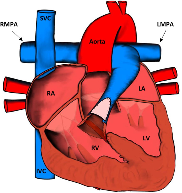
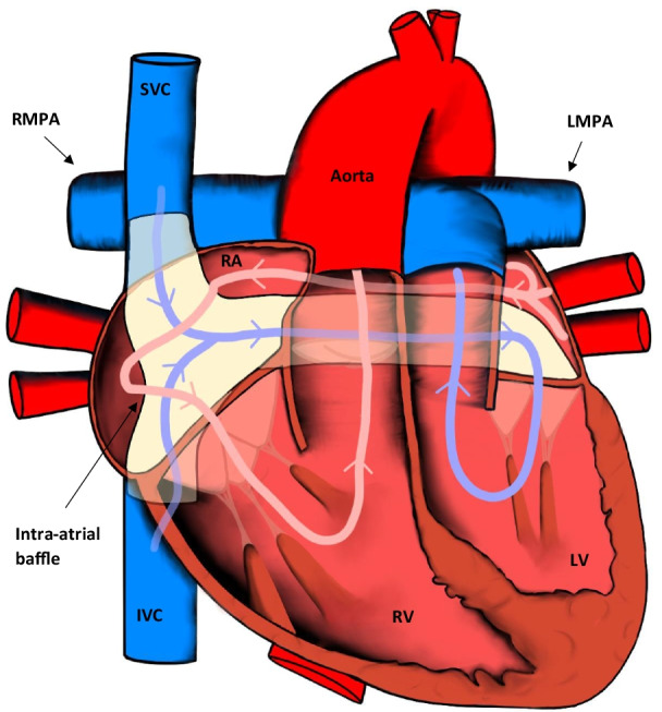
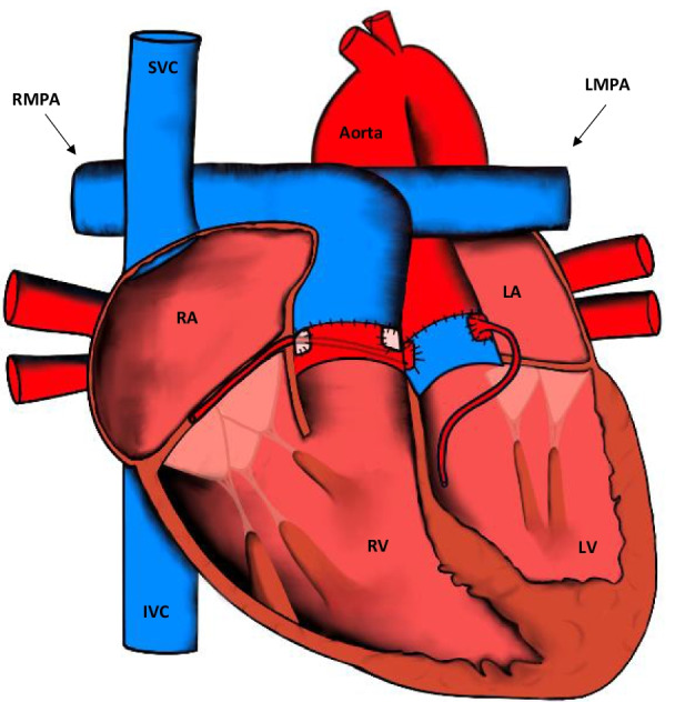
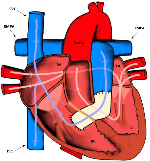
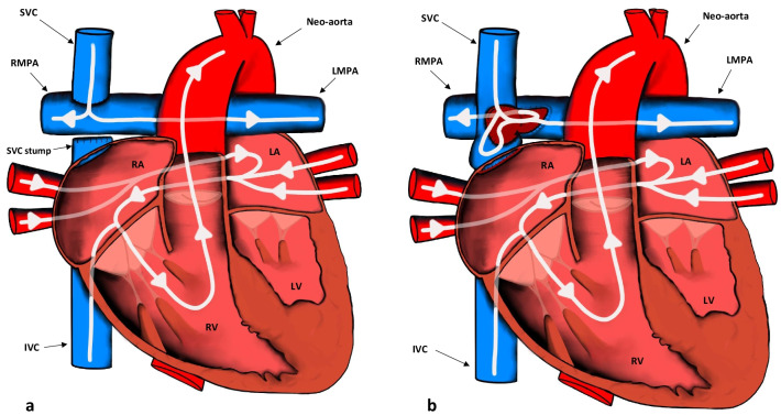
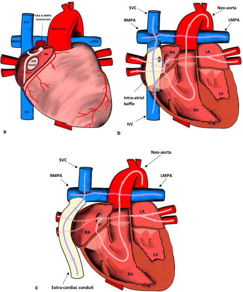
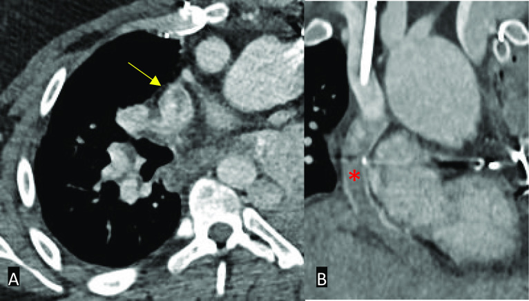
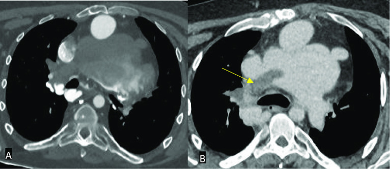
Congenital heart disease is the most common group of congenital pathology. Over the past few decades, advances in surgical treatment have resulted in a rising population of adult patients with repaired complex congenital heart disease. Although the quality of life has greatly improved, a significant proportion of morbidities encountered in clinical practice is now seen in adults rather than in children. These patients often have significant haemodynamic pathophysiology necessitating repeat intervention. CT and MRI are excellent imaging modalities, which help elucidate potential complications that may need urgent management. Although imaging should be performed in specialised centres, occasionally patients may present acutely to emergency departments in hospitals with little experience in managing potentially complex patients. The purpose of this article is to provide an introductory overview to the radiologist who may not be familiar with complex congenital heart disease in adult patients. This educational review has three main sections: (1) a brief overview of the post-operative anatomy and surgical management of the most common complex conditions followed by (2) a discussion on CT/MRI protocols and (3) a review of the various complications and their CT/MRI findings.

Keywords: Adult congenital heart disease; Computed tomography (CT); Magnetic resonance imaging (MRI).


Conflict of interest statement

The authors declare that they have no competing interests.

Figures

Fig. 1
Fig. 1
Appearances of the heart after TOF repair. The RVOT is repaired and the VSD is closed. IVC: inferior vena cava; LA: left atrium; LV: left ventricle; LMPA: left main pulmonary artery; RMPA: right main pulmonary artery; RA: right atrium; RV: right ventricle; SVC: superior vena cava
Fig. 2
Fig. 2
Appearances of the heart after the atrial switch procedure. The SVC and IVC are baffled to the LV, whereas the pulmonary veins are baffled to the RV. IVC: inferior vena cava; LV: left ventricle; LMPA: left main pulmonary artery; RMPA: right main pulmonary artery; RA: right atrium; RV: right ventricle; SVC: superior vena cava
Fig. 3
Fig. 3
Appearances of the heart after the arterial switch procedure. The aorta and the pulmonary trunk are transected near their origin and translocated along with the coronary arteries. IVC: inferior vena cava; LA: left atrium; LV: left ventricle; LMPA: left main pulmonary artery; RMPA: right main pulmonary artery; RA: right atrium; RV: right ventricle; SVC: superior vena cava
Fig. 4
Fig. 4
Appearances of the heart after the Rastelli procedure. The VSD is closed and an RV to pulmonary artery conduit is placed. IVC: inferior vena cava; LA: left atrium; LV: left ventricle; LMPA: left main pulmonary artery; RMPA: right main pulmonary artery; RA: right atrium; RV: right ventricle; SVC: superior vena cava
Fig. 5
Fig. 5
Appearances of the heart after the Norwood procedure (Stage 1), which involves construction of a neo-aorta, atrial septectomy, addition of a modified Blalock–Taussig shunt, and ligation of the ductus arteriosus. IVC: inferior vena cava; LA: left atrium; LV: left ventricle; LMPA: left main pulmonary artery; RMPA: right main pulmonary artery; RA: right atrium; RV: right ventricle; SVC: superior vena cava
Fig. 6
Fig. 6
a Appearances of the heart after the bi-directional Glenn procedure (Stage 2). Note that the SVC is closed off at or near its junction with the RA in contrast to the hemi-Fontan procedure. IVC: inferior vena cava; LA: left atrium; LV: left ventricle; LMPA: left main pulmonary artery; RMPA: right main pulmonary artery; RA: right atrium; RV: right ventricle; SVC: superior vena cava. b Appearances of the heart after the hemi-Fontan procedure (Stage 2). Note that the SVC remains in continuity with the RA in contrast to the bi-directional Glenn procedure. IVC: inferior vena cava; LA: left atrium; LV: left ventricle; LMPA: left main pulmonary artery; RMPA: right main pulmonary artery; RA: right atrium; RV: right ventricle; SVC: superior vena cava
Fig. 7
Fig. 7
a The last and final stage (Stage 3) for definitive repair of single ventricle defects. The IVC is connected to the RMPA to form the atriopulmonary variation of the Fontan procedure. The ASD is closed, and the RAA is anastomosed to the RMPA. ASD: atrial septal defect; IVC: inferior vena cava; LMPA: left main pulmonary artery; RAA: right atrial appendage; RMPA: right main pulmonary artery; RA: right atrium; SVC: superior vena cava. b The lateral intracardiac tunnel variation of the total cavopulmonary connection Fontan procedure. A baffle is placed inside the RA, and the SVC is connected directly to the RMPA. A fenestration to relieve pressure in the circuit may be seen between the baffle and the RA. IVC: inferior vena cava; LA: left atrium; LV: left ventricle; LMPA: left main pulmonary artery; RMPA: right main pulmonary artery; RA: right atrium; RV: right ventricle; SVC: superior vena cava. c The extracardiac conduit variation of the total cavopulmonary connection Fontan procedure. Note that the conduit that directs venous blood to the RMPA is extracardiac as opposed to the lateral intracardiac tunnel variation. LA: left atrium; LV: left ventricle; LMPA: left main pulmonary artery; RMPA: right main pulmonary artery; RA: right atrium; RV: right ventricle; SVC: superior vena cava
Fig. 8
Fig. 8
Axial (A) and coronal (B) contrast-enhanced CT images in a patient with an extra-cardiac conduit Fontan circuit. A If the study is protocolled as a standard CT pulmonary angiogram, a pseudo-thromboembolism may be seen – in this case seen in the superior vena cava (yellow arrow). B Note that the conduit also appears poorly opacified (red asterisk)
Fig. 9
Fig. 9
Axial CT images in a patient with an extra-cardiac conduit Fontan circuit. A Inadequate protocolling which resulted in suboptimal opacification of the circuit. Note that it is impossible to identify a true thrombus. B A single contrast phase, delayed phase acquisition protocol was subsequently used in the same patient which opacified the Fontan circuit adequately and allowed identification of a thrombus in the right main pulmonary artery (yellow arrow)
Fig. 10
Fig. 10
Four-chamber MRI SSFP cine in a patient post-TOF repair. There is significant right ventricular hypertrophy and dilatation (red asterisk). The interventricular septum is also shifted towards the left ventricle (yellow arrow)
Fig. 11
Fig. 11
Still shots of MRI SSFP cine in axial (A), coronal (B) planes and mid-ventricular short-axis views of inversion recovery sequence in the late gadolinium phase (C, D) demonstrating the appearances of the heart post-atrial switch procedure for D-TGA. A Normal appearances of the baffled pulmonary venous system to the right atrium and B venae cavae to the left atrium. C, D Right ventricular dilatation and hypertrophy with late gadolinium enhancement at the insertion points extending into the mid-septum (yellow arrows)
Fig. 12
Fig. 12
Select multiplanar CT images showing a valved RV-PA conduit to treat RVOT obstruction. Axial images in the bone window (A and C), lung window (B) and MPR reformats in the sagittal plane (D), bone window. There are vegetations of the RV-PA conduit (orange arrows), septic emboli in the arteries of the right lower lobe (blue arrow) and pulmonary abscesses (dotted circles). RV: right ventricle; PA: pulmonary artery; MPR: multiplanar reconstruction; RVOT: right ventricular outflow tract
Fig. 13
Fig. 13
Still shots of MRI SSFP cine in axial (A), sagittal oblique (B), coronal left ventricular outflow tract (C), and right ventricular inflow and outflow (D) views post-arterial switch for D-TGA. A Typical appearances of the Lecompte manoeuvre for the Jatene procedure. The pulmonary arteries bifurcate anterior to the ascending aorta, classically ‘draping’ over it. B Typical appearances of a high aortic arch. C Dilatation of the neo-aortic root (blue asterisk). D Normal appearances of the neo-pulmonary root (red asterisk). RPA: right pulmonary artery; LPA: left pulmonary artery
Fig. 14
Fig. 14
Select axial CT images in the bone (A and C), soft tissue (B), and lung windows (D). A Large perimembranous VSD (red asterisk) and right ventricular hypertrophy. B Dilated pulmonary trunk (blue asterisk) C reflux of contrast into the IVC and hepatic veins D bilateral pulmonary parenchymal mosaicism. Findings are compatible with pulmonary hypertension and right ventricular heart strain due to a long-standing VSD. Although this is an example of an isolated VSD, similar features of pulmonary hypertension can be seen in large residual VSDs in the setting of repaired TOF or repaired D-TGA with a VSD
Fig. 15
Fig. 15
A large right-sided pulmonary AVM in a patient with a Fontan circulation. a, b Axial still shots of MRI SSFP cine sequences demonstrating a pulmonary AVM in the right lower lobe, c, d catheter angiogram confirming the presence of an AVM, eh contrast-enhanced MRI angiogram in coronal planes demonstrating the AVM (white arrows). AVM: arteriovenous malformation. Re-used with permission from Fontan circulation in an adult: A guide for the radiologist. Arzanauskaite M, Nyktari E, Voges I. ESTI-ESCR 2018 / P-0103
Fig. 16
Fig. 16
A CT Axial image in the lung window in a patient with plastic bronchitis. Note the groundglass parenchymal lung changes in the right upper lobe. B A bronchial cast retrieved in a patient with plastic bronchitis
Fig. 17
Fig. 17
MRI SSFP cine (A) and contrast-enhanced CT (B) images in a patient with a valved (red asterisk) RV-PA conduit. A Single shot of the RVOT demonstrating pulmonary regurgitation (blue arrow). B Single axial image demonstrating calcification around the valve (yellow arrow). RV: right ventricle; PA: pulmonary artery; RVOT: right ventricular outflow tract
Fig. 18
Fig. 18
MRI SSFP cine images in two different patients post-TOF repair taken in the RVOT plane. A There is RVOT residual obstruction with a stenotic jet (blue arrow) with RV dilatation (red asterisk). B A jet of pulmonary regurgitation can be seen (yellow arrow). RVOT: right ventricular outflow tract; RV: right ventricle
Fig. 19
Fig. 19
Still shot post-contrast MR angiogram in the axial view. Residual right branch pulmonary stenosis after TOF repair. RPA: right pulmonary artery; LPA: left pulmonary artery (dilated)
Fig. 20
Fig. 20
Still-shot MRI SSFP cine images in the coronal (A) and sagittal (B) plane post-atrial switch for D-TGA. Baffle leak representing a large and broad tunnel from the IVC to the right atrium with a right-to-left shunt (Qp/Qs = 0.7). Dark blue asterisk: pulmonary venous channel; light blue asterisk: IVC channel; red arrow: baffle leak
Fig. 21
Fig. 21
Select coronary CT angiography images (A, B, C) in a patient post-coronary artery translocation. A Axial view showing ostial stenosis of the LCA (yellow arrow). B Reconstructed view of the LCA showing ostial stenosis (yellow arrow) and post-stenotic dilatation (red asterisk). C Axial view showing the normal ostium of the RCA (blue arrow) for comparison. D Catheter angiogram of the LCA showing ostial stenosis (yellow arrow). LCA: left coronary artery; RCA: right coronary artery
Fig. 22
Fig. 22
Still-shot MRI SSFP cine in the coronal view. Stenosis within the intra-atrial tunnel in a patient with a Fontan circuit (blue arrow). Re-used with permission from Fontan circulation in an adult: A guide for the radiologist. Arzanauskaite M, Nyktari E, Voges I. ESTI-ESCR 2018 / P-0103
Fig. 23
Fig. 23
ECG-gated CT study of an atriopulmonary Fontan circulation performed for congenital tricuspid atresia. Axial, coronal and sagittal views in the soft tissue window (AC): Large thrombus seen in the right atrium (blue arrows) adhered to the atrial wall. Re-used with permission from Fontan circulation in an adult: A guide for the radiologist. Arzanauskaite M, Nyktari E, Voges I. ESTI-ESCR 2018 / P-0103
Fig. 24
Fig. 24
Horizontal long-axis (A), sagittal (B, D), coronal (C), axial (E) and right pulmonary artery cross-cut views (F) of a cardiovascular MRI study in an atriopulmonary Fontan circulation performed for congenital tricuspid atresia. There are filling defects in keeping with thrombi in the right atrium (blue arrows) and in the right pulmonary artery (yellow arrows) on inversion recovery early-phase gadolinium sequences. Note that thrombus appears as very hypointense. Re-used with permission from Fontan circulation in an adult: A guide for the radiologist. Arzanauskaite M, Nyktari E, Voges I. ESTI-ESCR 2018/P-0103

References

    1. Gilboa SM, Devine OJ, Kucik JE, et al. Congenital heart defects in the United States: estimating the magnitude of the affected population in 2010. Circulation. 2016;134:101–109. doi: 10.1161/CIRCULATIONAHA.115.019307. - DOI - PMC - PubMed
    1. Stout KK, Daniels CJ, Aboulhosn JA, et al. 2018 AHA/ACC guideline for the management of adults with congenital heart disease: a report of the American College of Cardiology/American Heart Association task force on clinical practice guidelines. Circulation. 2019;139:e698–e800. - PubMed
    1. Sachdeva R, Valente AM, Armstrong AK, et al. ACC/AHA/ASE/HRS/ISACHD/SCAI/SCCT/SCMR/SOPE 2020 appropriate use criteria for multimodality imaging during the follow up care of patients with congenital heart disease: a report of the American College of Cardiology solution set oversight committee and appropriate use criteria task force, American Heart Association, American Society of Echocardiography, Heart Rhythm Society, International Society for Adult Congenital Heart Disease, Society for Cardiovascular Angiography and Interventions, Society of Cardiovascular Computed Tomography, Society for Cardiovascular Magnetic Resonance, and Society of Pediatric Echocardiography. J Am Coll Cardiol. 2020;75:657–703. doi: 10.1016/j.jacc.2019.10.002. - DOI - PubMed
    1. Burchill LJ, Huang J, Tretter JT, et al. Noninvasive imaging in adult congenital heart disease. Circ Res. 2017;120:995–1014. doi: 10.1161/CIRCRESAHA.116.308983. - DOI - PubMed
    1. Araujo JJ. The profile of an adult with congenital heart disease. Int J Clin Cardiol. 2018;5:131.

LinkOut - more resources