Review
. 2022 Apr 25;13(1):80.
doi: 10.1186/s13244-022-01218-3.

Radiology-pathology correlation of endometrial carcinoma assessment on magnetic resonance imaging

Affiliations
Review

Radiology-pathology correlation of endometrial carcinoma assessment on magnetic resonance imaging

Eveline Dokter et al. Insights Imaging. .

Abstract

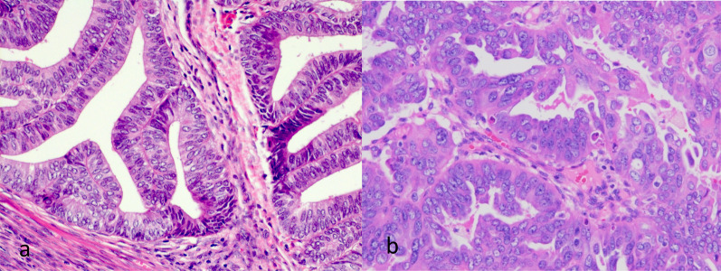
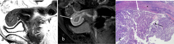
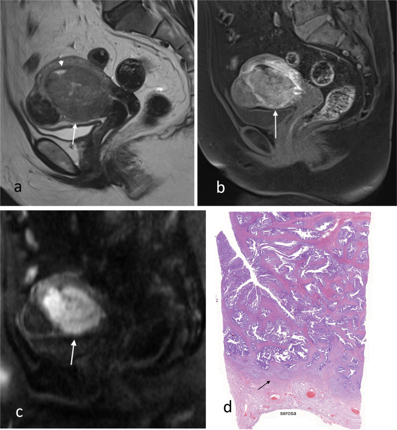
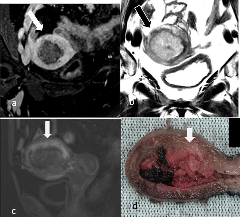
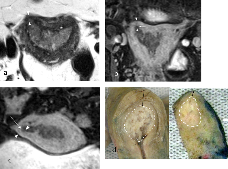
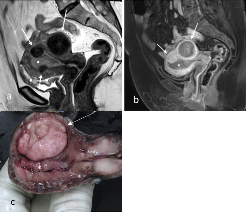
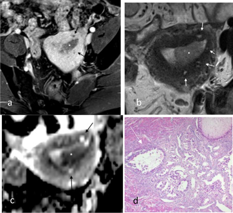
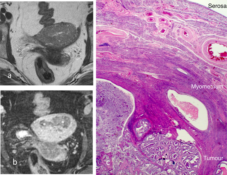
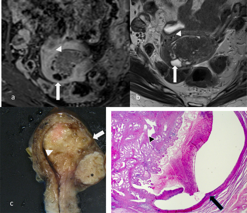
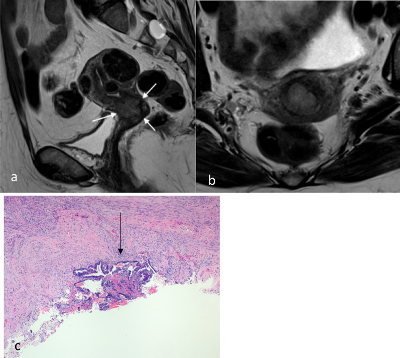
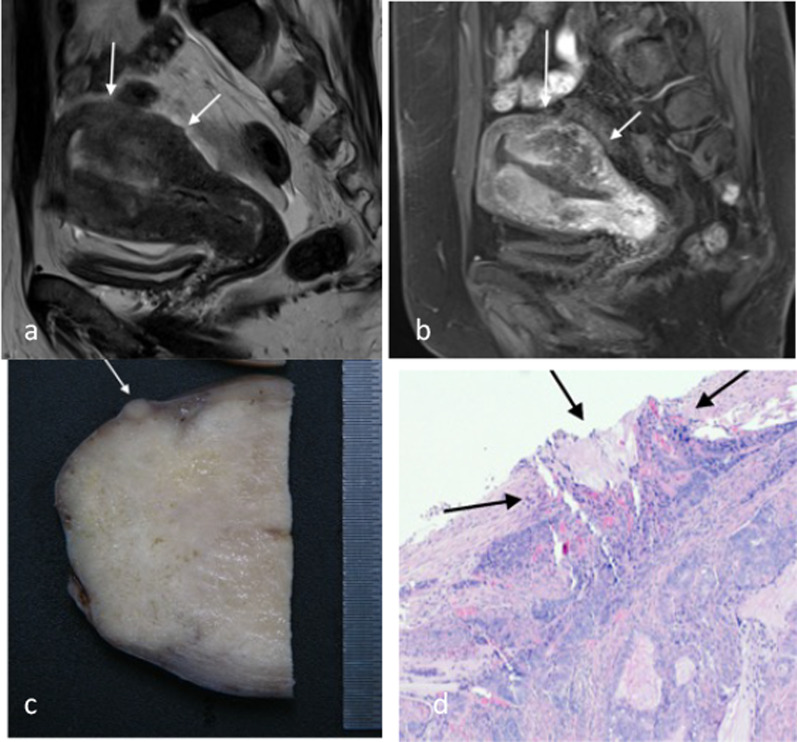
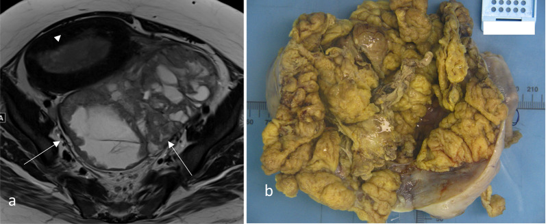
Endometrial carcinoma is the most common gynaecological cancer in developed countries. Most cases are low-volume/low-grade tumour at presentation; however, high-grade subtypes may present with locally advanced disease with higher propensity for spread outside of the pelvis. MRI has a role in local staging of the tumour and helping the clinicians in treatment decision making. This pictorial essay gives examples of endometrial carcinoma at different stages with histological correlation. It also explores the potential limitations and pitfalls of imaging in this context.

Keywords: Endometrial carcinoma; FIGO staging; Magnetic resonance imaging; Radiology-pathology correlation.


Conflict of interest statement

The authors declare that they have no competing interests.

Figures

Fig. 1
Fig. 1
Histology images of Type 1 (a) and Type 2 (b) endometrial carcinomas. a shows a grade 1 endometrioid adenocarcinoma. There are well-formed glands with uniform appearing pencillate nuclei. b shows a serous adenocarcinoma—a Type 2 tumour with increased propensity for early extra-uterine spread. There is a more haphazard and disorderly glandular growth with pale enlarged nuclei and prominent nucleoli
Fig. 2
Fig. 2
Stage 1A endometrioid endometrial carcinoma without myometrial invasion. Sagittal T2W image (a) shows the tumour is intermediate in signal (arrow). On early sagittal DCE image (b), there is smooth uninterrupted band of subendometrial junctional zone enhancement (arrow) which excludes myometrial invasion. Histology image (c) shows tumour (black arrow) confined to the endometrium and does not invade into the myometrium (*). There is preserved endometrium/myometrium junction (white arrow)
Fig. 3
Fig. 3
Stage IB endometrial carcinoma with extension to the outer half of the myometrium. Sagittal T2W image (a) shows the tumour (arrow) is bulky and extends into the outer half of the myometrium anteriorly. The outer uterine contour remains smooth. Junctional zone posteriorly is preserved as a thin T2 hypointense line (arrowhead). Sagittal early DCE images (b) show irregular peritumoural enhancement at the advancing front of the tumour (arrow) anteriorly, whilst the normal junctional zone posteriorly (arrowhead) has a smooth thin enhancement. Sagittal DW image (c) shows marked diffusion restriction of the tumour (arrow). Low-power histology (d) shows tumour extension to the outer half of the myometrium with preserved serosa and small amount of normal residual myometrium (arrow)
Fig. 4
Fig. 4
Radiological (a, oblique short-axis post-contrast T1W GRE fat suppressed image) and pathological (b, 0.4 × magnification) measurement of depth of myometrial invasion. (a) shows the radiological measurement of depth of invasion. The red line is an imaginary line estimating the endometrium/myometrium junction. Measurement (a arrow) shows the depth of the tumour into the myometrium beyond the junction. Measurement (b arrow) shows overall total thickness of the myometrium. The endometrium/myometrium junction can be irregular normally or due to distortion by the tumour. Measurement on histology is performed on low-power view with myoinvasion determined when tumour cell is seen interspersed with myocytes. The solid line delineates the normal endometrium from the myometrium. The dotted line is the estimated endometrium/myometrium junction at the site of the tumour and the basis for the measurement of myoinvasion
Fig. 5
Fig. 5
Oblique axial post-contrast 3D T1W GRE MR (a), oblique axial T2W MR (b), sagittal early phase DCE T1W MR (c) and macroscopic (d) images of a large endometrial carcinoma. There is a large exophytic intracavitary tumour with distension of the endometrial cavity. In this situation, it is difficult to estimate where the endometrium/myometrium interface should be and the depth of invasion. In this case, the tumour was extending into the outer half of the myometrium (stage 1b) at the posterior aspect on final pathology. The invasive front of the tumour (arrow) shows early peritumoural enhancement and has slight irregularity at the interface with the normal myometrium and blurring of junctional zone’s low T2 signal
Fig. 6
Fig. 6
Tumour extending to the right cornua in a patient with stage 1a endometrial carcinoma. Axial T2W image (a), oblique coronal (b) and oblique axial (c) post-contrast 3D T1W GRE fat suppressed reformat images show the tumour involving the endometrial cavity and extending to the right cornu (arrow). There is normal thinning of the myometrium (arrowheads) at the cornu compared to the background myometrium. Midline sagittal and right cornu parasagittal macroscopic specimens (d) show the tumour (dotted white lines) and thinning of the myometrium (arrows) at the cornu. The background myometrial thinning has to be taken into account in the evaluation of the percentage of the depth of invasion to avoid overestimation
Fig. 7
Fig. 7
Sagittal T2W (a), sagittal post-contrast T1W (b) and macroscopic (c) images of stage 1a endometrial carcinoma (*) without myometrial invasion in a multi-fibroid uterus. The fibroids (arrows) are T2 hypointense and often hypoenhancing relative to the myometrium. The fibroids distort the endometrial–myometrial junction and complicate the assessment for myometrial invasion. The fundal fibroid shows similar enhancement relative to the myometrium
Fig. 8
Fig. 8
Endometrial carcinoma in a patient with adenomyosis. Axial post-contrast T1W GRE (a), axial T2W (b) and axial ADC map (c) images show the tumour (*) in the endometrial cavity. There is diffuse adenomyosis with background myometrial cysts (arrows). Myometrial cysts are hyperintense on T2 weighted imaging without significant diffusion restriction. Tumour can colonise adenomyosis and is not considered to be muscle invasive if confined to the adenomyosis. Histology (H&E stain, 4 × magnification, d) showed colonisation of areas of adenomyosis by complex crowded tumour cells (arrow), but this could not be preoperatively distinguished from normal adenomyosis on imaging
Fig. 9
Fig. 9
Endometrial carcinoma with T2 signal isointense to myometrium. On axial T2W MR image (a), the tumour has the same signal intensity to the adjacent myometrium. It is difficult to appreciate the exact border on T2W MR imaging. On the post-contrast T1W imaging (b), there is better tumour (*) to myometrium contrast. Microscopic image (c) confirms tumour invasion into the myometrium
Fig. 10
Fig. 10
Endometrial carcinoma with mucinous differentiation. On the oblique axial post-contrast T1W (a) and the T2W (b) MR images, the tumour is accompanied by cystic spaces that are T2 hyperintense without enhancement (arrows). These cystic areas on pathology (c) correlate with mucin pools (arrow) associated with the tumour (arrowheads) that extend into the outer half of the myometrium. There is a fibroid in the lower aspect of the uterus (*). Microscopic image (d) shows endometrioid carcinoma (arrowhead) with mucinous differentiation and large space adjacent (arrow) containing acellular proteinaceous material consistent with mucin
Fig. 11
Fig. 11
Stage II endometrial carcinoma with cervical stroma involvement. On sagittal (a) and axial (b) T2W MR imaging, there is intermediate T2 signal tumour (arrows) replacing the low signal dense cervical stromal tissue. In this case, the tumour involving the cervix is from a drop metastasis from a smaller primary in the endometrial cavity (not shown). Pathology (c) determines cervical invasion based on identification of the haphazardly arranged tumour cells (arrow) invading beyond the confines of cervical mucosa with surrounding stromal reaction
Fig. 12
Fig. 12
Stage IIIA endometrial carcinoma with serosal involvement. There is irregularity at the posterior uterine outer surface (arrows) on sagittal T2W (a) and sagittal DCE T1 MR imaging (b) in addition to full thickness tumour signal involvement of the myometrium. Pathological examples of serosal involvement are shown in c, d. Macroscopically, there is a homogeneous tan nodule at the serosal surface (arrow, c). Microscopically, this correlates with infiltrating hyperchromatic tumour cells extending through the outer myometrium and erupting through the serosal tissue (arrow, d)
Fig. 13
Fig. 13
Stage IIIA endometrial carcinoma. Axial T2W MR image (a) shows a left ovarian metastasis (arrow) separate from the primary tumour (arrowhead). Macroscopic pathology specimen (b) shows open ovary with internal friable and tan solid components. Ovarian involvement can be due to direct invasion, haematogenous spread or transcoelomic spread resulting in serosal tumour deposits
Fig. 14
Fig. 14
Serosal deposit in ovarian parenchyma. Histology image (H&E stain, 2 × magnification) shows a small serosal deposit in the ovarian parenchyma. This was not visible on MRI. The small volume of disease may account for the lower sensitivity of MRI for ovarian metastases in endometrial carcinoma
Fig. 15
Fig. 15
Stage IIIB endometrial carcinoma with vaginal involvement. There is a large tumour with extension through the myometrium into the serosa. On the sagittal (a) and axial (b) T2W MR images, there is a nodule (arrow) with intermediate T2 signal caudally at the level of the vagina on the right, in keeping with a drop metastasis. On axial post-contrast T1W image (c), the drop metastasis is an hypoenhancing lesion (arrow) compared to the hyperenhancing vaginal mucosa and muscular layer. This was diagnosed with biopsies confirming endometrioid carcinoma
Fig. 16
Fig. 16
Stage IIIB endometrial carcinoma with parametrial invasion. Coronal T2W MR image (a) shows irregular intermediate T2 tumour signal replacing the entirety of the cervix and extending into the left parametrial fat in keeping with parametrial invasion (arrow). Parametrial invasion occurs from contiguous spread via the cervix. On histological image (× 10 magnification) (b), this correlates with the presence of tumour cells (arrowheads) infiltrating into adjacent adipose tissue (arrow) with a desmoplastic stromal response (*)
Fig. 17
Fig. 17
Isolated tumour cells less than 0.2 mm diameter in a lymph node detected on AE1/AE3 cytokeratin immunohistochemistry stain (× 20 magnification). Such low-volume disease in nodal spread may account for MRI’s limited sensitivity for lymph node involvement
Fig. 18
Fig. 18
Stage IVA endometrial carcinoma. Sagittal T2W MR images (a) show direct tumour extension into the bladder mucosa (arrow). The air anteriorly in the bladder is due to a fistula. Chips of a bladder tumour (b, H&E stain, × 4 magnification) initially thought to be primary urothelial tumour show fragments of necrotic endometrioid adenocarcinoma (arrow) confirmed on immunohistochemistry. The normal bladder transitional epithelium is indicated by the arrowheads

References

    1. Brand A, Hammond I, Cheuk R et al (2020) Clinical practice guidelines for the treatment and management of endometrial cancer. Cancer Council
    1. Siegel RL, Miller KD, Jemal A. Cancer statistics, 2020. CA. 2020;70(1):7–30. doi: 10.3322/caac.21590. - DOI - PubMed
    1. Murali R, Soslow RA, Weigelt B. Classification of endometrial carcinoma: more than two types. Lancet Oncol. 2014;15(7):e268–278. doi: 10.1016/S1470-2045(13)70591-6. - DOI - PubMed
    1. Rauch G, Kaur H, Choi H, Ernst R, Klopp A, Boonsirikamchai P, Westin S, Marcal L. Optimization of MR imaging for pretreatment evaluation of patients with endometrial and cervical cancer. Radiographics. 2014;34:1082–1098. doi: 10.1148/rg.344140001. - DOI - PMC - PubMed
    1. Otero-Garcia MM, Mesa-Alvarez A, Nikolic O, Blanco-Lobato P, Basta-Nikolic M, de Llano-Ortega RM, Paredes-Velazquez L, Nikolic N, Szewczyk-Bieda M. Role of MRI in staging and follow-up of endometrial and cervical cancer: pitfalls and mimickers. Insights Imaging. 2019;10(1):19. doi: 10.1186/s13244-019-0696-8. - DOI - PMC - PubMed

LinkOut - more resources