. 2022 Apr 7:(182):10.3791/63539.
doi: 10.3791/63539.

Echocardiographic Characterization of Left Ventricular Structure, Function, and Coronary Flow in Neonate Mice

Affiliations

Echocardiographic Characterization of Left Ventricular Structure, Function, and Coronary Flow in Neonate Mice

Shamim A K Chowdhury et al. J Vis Exp. .

Abstract

Echocardiography is a non-invasive procedure that enables the evaluation of structural and functional parameters in animal models of cardiovascular disease and is used to assess the impact of potential treatments in preclinical studies. Echocardiographic studies are usually conducted in young adult mice (i.e., 4-6 weeks of age). The evaluation of early neonatal cardiovascular function is not usually performed because of the small size of the mouse pups and the associated technical difficulties. One of the most important challenges is that the short length of the pups' limbs prevents them from reaching the electrodes in the echocardiography platform. Body temperature is the other challenge, as pups are very susceptible to changes in temperature. Therefore, it is important to establish a practical guide for performing echocardiographic studies in small mouse pups to help researchers detect early pathological changes and study the progression of cardiovascular disease over time. The current work describes a protocol for performing echocardiography in mouse pups at the early age of 7 days old. The echocardiographic characterization of cardiac morphology, function, and coronary flow in neonatal mice is also described.


Conflict of interest statement

Disclosures

The authors have nothing to disclose.

Figures
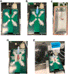
Figure 1:
Figure 1:. Echocardiographic platform setup and 7-day-old mouse pup preparation.
(A) Aluminum foil strips are placed on the platform electrode pads and secured with tape. (B) The glove finger is cut and adapted to fit the isoflurane/oxygen nose cone. (C) The pup is placed in the isoflurane induction chamber, and the isoflurane delivery starts at 1.5% concentration. (D) The pup is placed in a supine position with paws touching the aluminum foil strips and secured with tape. Two rolls of gauze are used to keep the acoustic gel in place. (E) A heating lamp is placed close to the pup to maintain its body temperature.
Figure 2:
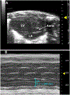
Figure 2:. Parasternal long-axis (PLAX) view of the left ventricle.
(A) B-mode images of the left ventricular chamber (LV), left atrium (LA), and the aorta. (B) M-mode is used to measure the LA diameter.
Figure 3:
Figure 3:. Parasternal short-axis (PSAX) view of the left ventricle.
(A) B-mode images of the left ventricular chamber. (B) M-mode sample of the interventricular septum at diastole (IVSd), left ventricular internal diameter at diastole (LVIDd), and posterior wall thickness at diastole (PWd). (C) Representative images of the pulmonary peak flow velocity, pulmonary ejection time (PET), and pulmonary acceleration time (PAT). (D) Representative images of the aortic ejection time (AET).
Figure 4:
Figure 4:. Apical four-chamber view.
(A) B-mode image of the left ventricle (LV), right ventricle (RV), left atrium (LA), and right atrium (RA). (B) Representative images of the maximal blood inflow velocity in the early phase of diastole (E), maximal blood inflow velocity in the late phase of diastole (A), deceleration time (DT), isovolumetric contraction time (IVCT), and isovolumetric relaxation time (IVRT). (C) Tissue Doppler sample images of the peak myocardial relaxation velocity in the early diastolic filling (e’), late diastolic filling (a’), and peak systolic myocardial velocity (s’).
Figure 5:
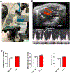
Figure 5:. Modified parasternal long-axis view.
(A) Platform and transducer position in modified parasternal long-axis view. (B) Visualization and recording of left anterior descending (LAD) coronary artery flow. LVOT = left ventricular outflow tract. (C) Peak coronary flow velocity (CFV), mean CFV, and velocity-time integral (VTI) in diastole are measured at 1.5% isoflurane (baseline) and 5 min after increasing isoflurane concentration to 2.5%; 7-day old mice, N = 7; data presented as mean ± SD.

References

    1. Le VP, Wagenseil JE Echocardiographic Characterization of Postnatal Development in Mice with Reduced Arterial Elasticity. Cardiovascular Engineering and Technology. 3 (4), 424–438 (2012). - PMC - PubMed
    1. Nagueh SF et al. Recommendations for the Evaluation of Left Ventricular Diastolic Function by Echocardiography: An Update from the American Society of Echocardiography and the European Association of Cardiovascular Imaging. European Heart Journal: Cardiovascular Imaging. 17 (12), 1321–1360 (2016). - PubMed
    1. Lang RM et al. Recommendations for cardiac chamber quantification by echocardiography in adults: an update from the American Society of Echocardiography and the European Association of Cardiovascular Imaging. European Heart Journal: Cardiovascular Imaging. 16 (3), 233–270 (2015). - PubMed
    1. Chrysant SG A new paradigm in the treatment of the cardiovascular disease continuum: focus on prevention. Hippokratia. 15 (1), 7–11 (2011). - PMC - PubMed
    1. Blom JN, Lu X, Arnold P, Feng Q Myocardial infarction in neonatal mice, a model of cardiac regeneration. Journal of Visualized Experiments. 111, e54100 (2016). - PMC - PubMed

Publication types

LinkOut - more resources