. 2022 Apr 25;14(8):3540-3553.
doi: 10.18632/aging.204028. Epub 2022 Apr 25.

MicroRNA-181a-5p prevents the progression of esophageal squamous cell carcinoma in vivo and in vitro via the MEK1-mediated ERK-MMP signaling pathway

Affiliations

MicroRNA-181a-5p prevents the progression of esophageal squamous cell carcinoma in vivo and in vitro via the MEK1-mediated ERK-MMP signaling pathway

Mingbo Wang et al. Aging (Albany NY). .

Abstract

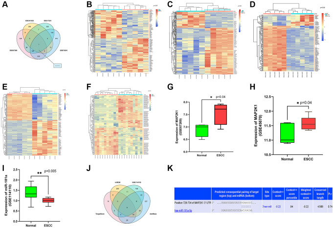
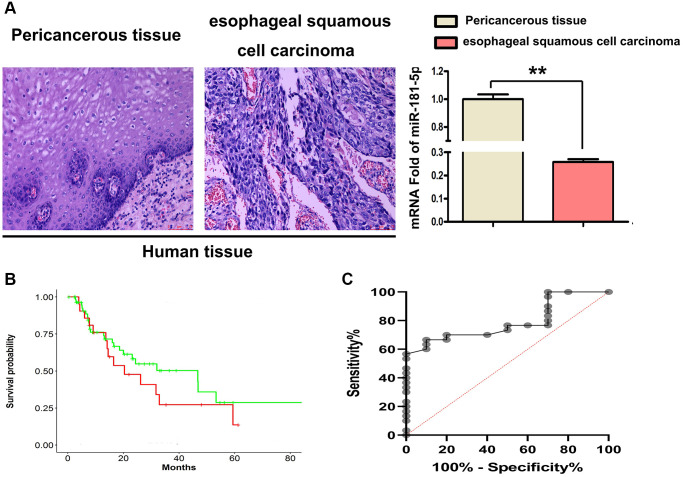
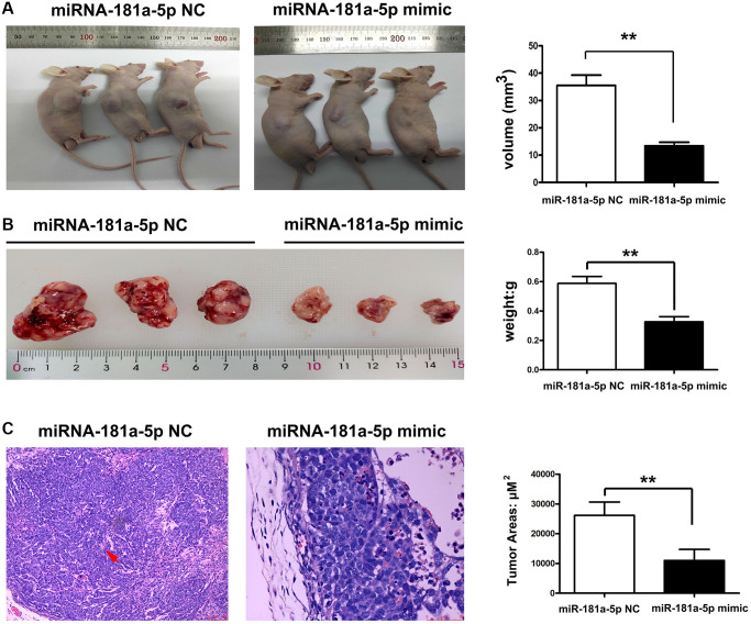
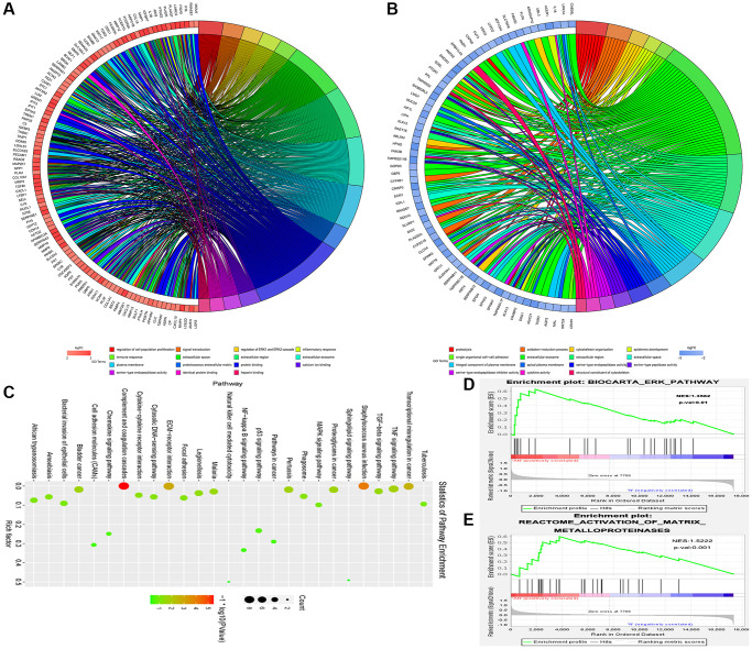
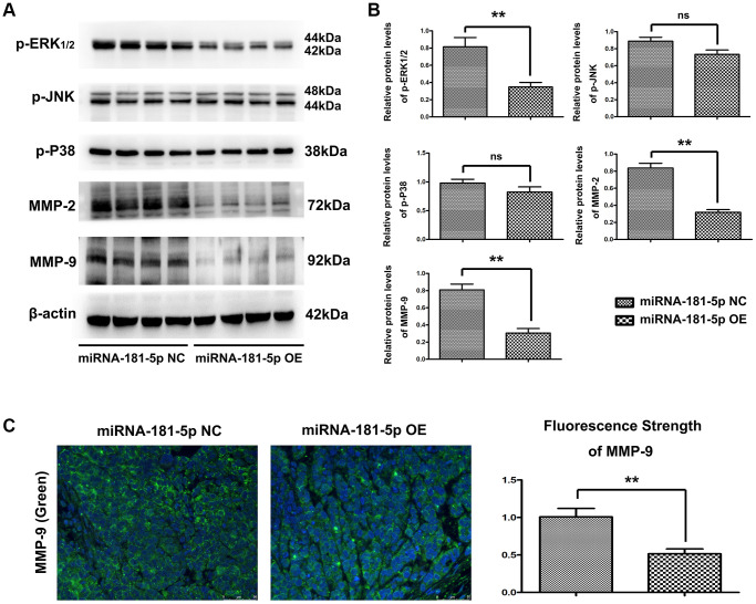
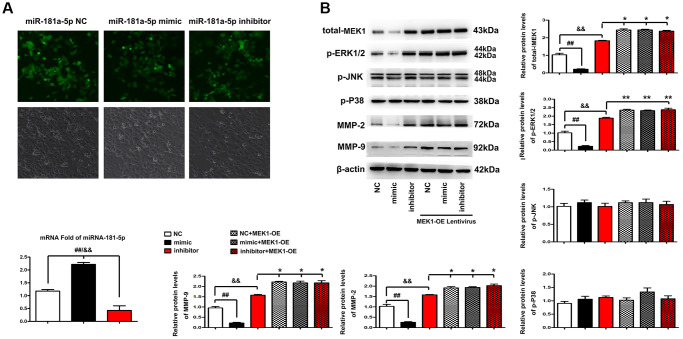
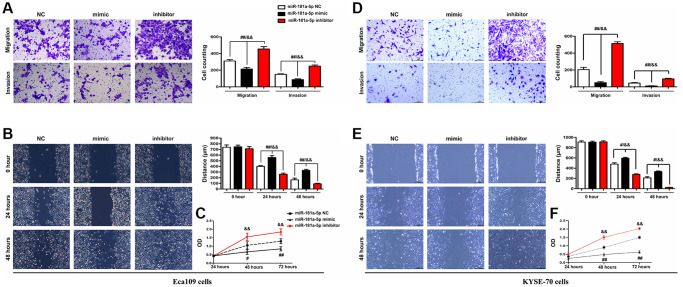
MicroRNAs (miRNAs) have been revealed to play a crucial role in oncogenesis of esophageal squamous cell carcinoma (ESCC). However, the biological role of miR-181a-5p in ESCC is currently less explored. The current study was designed to assess whether miR-181a-5p affects ESCC progression and further investigate relevant underlying mechanisms. Based on the data of GSE161533, GSE17351, GSE75241 and GSE67269 downloaded from GEO database, MAP2K1 (MEK1) was revealed to be one overlapping gene of the top 300 DGEs. Additionally, using the predicting software, miR-181a-5p was projected as the presumed target miRNA. Immunohistochemical staining and RT-qPCR research revealed that miR-181a-5p expression was decreased in human tumor tissues relative to surrounding peri-cancerous tissues. In an in vivo experiment, miR-181a-5p mimics could inhibit tumor growth and metastasis of ESCC. Gene expression profiles in combination with gene ontology (GO) and KEGG pathway analysis revealed that MAP2K1 (MEK1) gene and ERK-MMP pathway were implicated in ESCC progression. MiR-181a-5p mimics inhibited the activity of p-ERK1/2, MMP2 and MMP9 in vivo, as shown by Western blotting and immunohistochemistry labeling. There were no variations in the expression of p-P38 and p-JNK proteins. Additionally, miR-181a-5p mimics lowered p-ERK1/2, MMP2 and MMP9 levels in ECA109 cells, which were restored by MEK1-OE lentivirus. MEK1-OE Lentivirus significantly reversed the function induced by miR-181a-5p mimics in ECA109 cells. Moreover, further investigation indicated that the capability of migration, invasion and proliferation was repressed by miR-181a-5p mimics in ECA109 cells. In short, repressed ERK-MMP pathway mediated by miR-181a-5p can inhibit cell migration, invasion and proliferation by targeting MAP2K1 (MEK1) in ESCC.

Keywords: ESCC; MEK1; miR-181a-5p.


Conflict of interest statement

CONFLICTS OF INTEREST: The authors declare no conflicts of interest related to this study.

Figures

Figure 1
Figure 1
MiR-181a-5p was predicted as an applicant miRNA that affected the development of ESCC by targeting MAP2K1 (MEK1). (A) The top 300 DEGs determined from GSE161533, GSE17351, GSE75241 and GSE67269 datasets; (BE) The heat map depicted the top 100 DEGs from GSE161533, GSE17351, GSE75241 and GSE67269 datasets; (F) The heat map depicted the top 100 DEMs from GSE114110 (G) the expression of MAP2K1 in ESCC in the GSE67269 dataset; (H) the expression of MAP2K1 in ESCC in the GSE45670 dataset; (I) the expression of miR-181a in ESCC in the GSE114110 dataset; (J) the top 100 miRNAs predicted from TargetScan, miRDB, and starBase. (K) miR-181a-5p was found had targeted binding regions with MAP2K1 (MEK1).
Figure 2
Figure 2
MiR-181a-5p was downregulated in 4 paired human ESCC tissues. (A) H&E staining was used to identified ESCC tissues and their corresponding adjacent peri-cancerous tissue specimens, RT-qPCR of miR-181a-5p expression in human ESCC tissues. *P < 0.05, esophageal squamous cell carcinoma vs. peri-cancerous tissue. (B) Kaplan-Meier's method was used for survival analysis and miR-181 survival curve was drawn. (C) Kaplan-Meier analysis was used to depict the overall survival curves of patients with of ESCC from the data in GSE114110.
Figure 3
Figure 3
Upregulation of miR-181a-5p inhibited tumor growth and metastasis of ESCC. Nude mice were injected subcutaneously with ESCC cells and intratumorally injected with miR-181a-5p via. (A) The tumor volume in miR-181a-5p mimics and negative control mice; (B) the tumor weight in miR-181a-5p mimics and negative control mice; *P < 0.05 miR-181a-5p mimics vs. the NC groups; (C) H&E staining in experimental mice; *P < 0.05, **P < 0.01 miR-181a-5p mimics vs. the NC groups.
Figure 4
Figure 4
The MAP2K1 (MEK1) gene and ERK-MMP pathway were implicated in ESCC progression. Gene expression profiles in combination with gene ontology (GO) and KEGG pathway analysis was performed; (A) The results of the Go pathways from the data in GSE161533 dataset; (B) the results of the Go pathways from the data in GSE17351; (C) partial results of the KEGG pathways from the data in GSE17351; (D and E) the activation of matrix metalloproteinases signaling pathway and ERK signaling pathway were enriched in ESCC and appeared to be strongly correlated to with MEK1.
Figure 5
Figure 5
Elevation of miR-181a-5p reduced p-ERK, MMP2 and MMP9 in vivo. Nude mice were injected subcutaneously with ESCC cells and intratumorally injected with miR-181a-5p; (A and B) the protein expression of p-ERK1/2, p-JNK, p-P38, MMP2 and MMP9 as evaluated by Western blotting and RT-qPCR. *P < 0.05 miR-181a-5p mimics vs. NC group; (C) Immunohistochemical staining of MMP9 in miR-181a-5p mimics group and in negative control group; the positive expression rate of MMP9 in miR-181a-5p mimics group and negative control group. *P < 0.05, **P < 0.01 miR-181a-5p mimics vs. NC group.
Figure 6
Figure 6
Elevation of miR-181a-5p regulated ERK-MMP pathway in ESCC through MEK1. (A) FISH was performed to identify the distribution and expression levels of miR-181a-5p in miR-181a-5p NC, miR-181a-5p mimics or inhibitor transfected ECA109 cells. The positive expression shows green fluorescence. The red arrows represent a tiny amount of fluorescence of miR-181a-5p. (B) Western blotting analysis of p-ERK1/2, p-JNK, p-P38, t-ERK1, MMP2 and MMP9 in miR-181a-5p NC, miR-181a-5p mimics or inhibitor transfected ECA109 cells. The p-ERK1/2, p-JNK, p-P38, t-ERK1, MMP2 and MMP9 protein levels were measured after co-transfection with miR-181a-5p mimics, miR-181a-5p NC or miR-181a-5p inhibitor and MEK1-OE Lentivirus in ECA109 cells. Different data were presented as mean ± S.D. (all P < 0.05).
Figure 7
Figure 7
Elevation of miR-181a-5p repressed ESCC cell migration, invasion and proliferation. ECA109 and KYSE-70 cells were transfected with miR-181a-5p mimic, inhibitor or negative control (NC) for 48 hours; (A, D) Transwell invasion assay exposed that miR-181a-5p mimic inhibited cell migration and invasion in Eca109 (A) and KYSE-70 cells (B) Wound healing assay exposed that miR-181a-5p mimic inhibited cell migration. Representative images were obtained at 0, 24 and 48 h. RT-qPCR analysis of wound area in Eca109 (C) and KYSE-70 cells (D). The proliferation of ECA109 cells treated with miR-181a-5p mimic, inhibitor or negative control (NC) was measured by CCK8 assay in Eca109 (E) and KYSE-70 cells (F). *P < 0.05, **P < 0.01 miR-181a-5p mimics vs. NC group.

Similar articles

Cited by

References

    1. Xu CQ, Zhu ST, Wang M, Guo SL, Sun XJ, Cheng R, Xing J, Wang WH, Shao LL, Zhang ST. Pathway analysis of differentially expressed genes in human esophageal squamous cell carcinoma. Eur Rev Med Pharmacol Sci. 2015; 19:1652–61. - PubMed
    1. Chen YB, Jia WH. A comprehensive genomic characterization of esophageal squamous cell carcinoma: from prognostic analysis to in vivo assay. Chin J Cancer. 2016; 35:76. 10.1186/s40880-016-0142-y - DOI - PMC - PubMed
    1. Natsugoe S, Mueller J, Stein HJ, Feith M, Höfler H, Siewert JR. Micrometastasis and tumor cell microinvolvement of lymph nodes from esophageal squamous cell carcinoma: frequency, associated tumor characteristics, and impact on prognosis. Cancer. 1998; 83:858–66. 10.1002/(SICI)1097-0142(19980901)83:5<858::AID-CNCR9>3.0.CO;2-E - DOI - PubMed
    1. Zhao S, Jiang Y, Zhao J, Li H, Yin X, Wang Y, Xie Y, Chen X, Lu J, Dong Z, Liu K. Quercetin-3-methyl ether inhibits esophageal carcinogenesis by targeting the AKT/mTOR/p70S6K and MAPK pathways. Mol Carcinog. 2018; 57:1540–52. 10.1002/mc.22876 - DOI - PubMed
    1. Wang C, Fu X, Cai X, Wu X, Hu X, Fan M, Xiang J, Zhang Y, Chen H, Jiang G, Zhao K. High-dose nimotuzumab improves the survival rate of esophageal cancer patients who underwent radiotherapy. Onco Targets Ther. 2015; 9:117–22. 10.2147/OTT.S89592 - DOI - PMC - PubMed

MeSH terms