Observational Study
. 2022 Jul 5;15(7):423-434.
doi: 10.1158/1940-6207.CAPR-21-0610.

Chronic Exposure to Waterpipe Smoke Elicits Immunomodulatory and Carcinogenic Effects in the Lung

Affiliations
Observational Study

Chronic Exposure to Waterpipe Smoke Elicits Immunomodulatory and Carcinogenic Effects in the Lung

Maya Hassane et al. Cancer Prev Res (Phila). .

Abstract

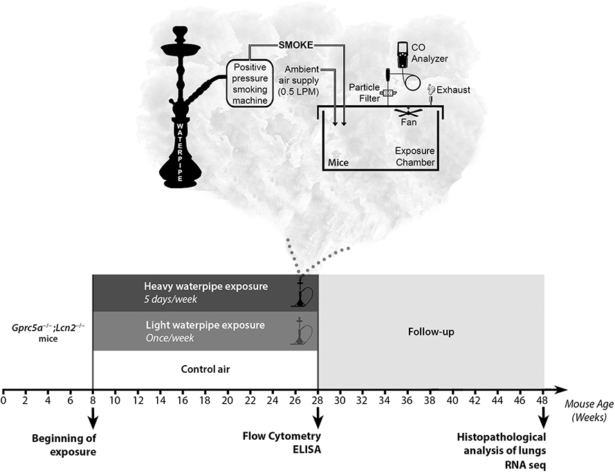
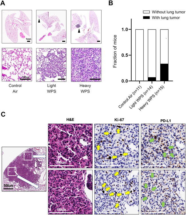
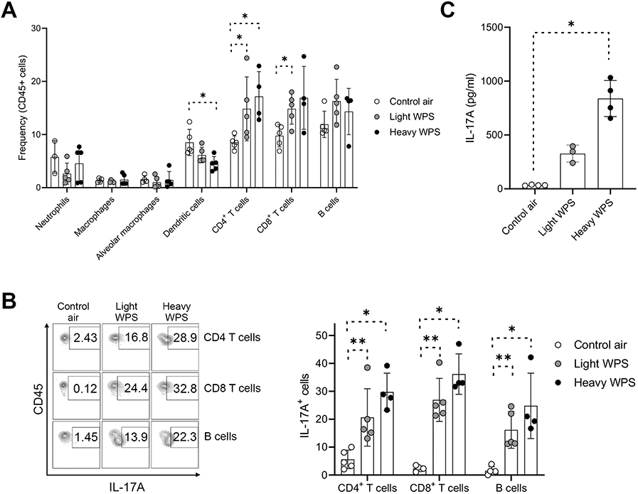
Effects of waterpipe smoking on lung pathobiology and carcinogenesis remain sparse despite the worldwide emergence of this tobacco vector. To address this gap, we investigated the effects of chronic waterpipe smoke (WPS) exposure on lung pathobiology, host immunity, and tumorigenesis using an experimental animal model that is prone to tobacco carcinogens and an exploratory observational analysis of human waterpipe smokers and nonsmokers. Mice exhibited elevated incidence of lung tumors following heavy WPS exposure (5 days/week for 20 weeks) compared to littermates with light WPS (once/week for 20 weeks) or control air. Lungs of mice exposed to heavy WPS showed augmented CD8+ and CD4+ T cell counts along with elevated protumor immune phenotypes including increased IL17A in T/B cells, PD-L1 on tumor and immune cells, and the proinflammatory cytokine IL1β in myeloid cells. RNA-sequencing (RNA-seq) analysis showed reduced antitumor immune gene signatures in animals exposed to heavy WPS relative to control air. We also performed RNA-seq analysis of airway epithelia from bronchial brushings of cancer-free waterpipe smokers and nonsmokers undergoing diagnostic bronchoscopy. Transcriptomes of normal airway cells in waterpipe smokers, relative to waterpipe nonsmokers, harbored gene programs that were associated with poor clinical outcomes in patients with lung adenocarcinoma, alluding to a WPS-associated molecular injury, like that established in response to cigarette smoking. Our findings support the notion that WPS exhibits carcinogenic effects and constitutes a possible risk factor for lung cancer as well as warrant future studies that can guide evidence-based policies for mitigating waterpipe smoking.

Prevention relevance: Potential carcinogenic effects of waterpipe smoking are very poorly understood despite its emergence as a socially acceptable form of smoking. Our work highlights carcinogenic effects of waterpipe smoking in the lung and, thus, accentuate the need for inclusion of individuals with exclusive waterpipe smoking in prevention and smoking cessation studies.


Conflict of interest statement

Competing interests: HK reports funding from Johnson and Johnson outside the scope of this work. All other authors declare no potential conflicts of interest.

Figures

Figure 1.
Figure 1.. Experimental design and waterpipe exposure apparatus.
Schematic design depicting the whole body waterpipe smoke exposure system. Smoke is drawn intermittently by a positive pressure smoking machine and discharged into a transparent polycarbonate chamber (38x25x25 cm) containing the animals. During the exposure event, ambient air is continuously piped into the chamber at a flow rate of 0.5 LPM. A CO analyzer (Bacharach Monoxor III) is used to monitor the CO levels in the chamber, and a fan is suspended to ensure uniform aerosol concentrations throughout the chamber. A glass fiber filter (Gelman Type A/E) is placed upstream of the CO monitor to allow gravimetric determination of the mean total particulate matter concentration during each exposure session. For the control group, the smoking machine remains off during each exposure session, and animals are exposed to ambient air only. Lower panel. Schematic timeline depicting the three experimental exposure groups composed of eight weeks-old Gprc5a−/−;Lcn2−/− mice divided into groups of up to five mice (per exposure group) and studied at the end of exposure, and 20 weeks after completion of exposure. Mice were exposed to WPS for once a week (light WPS) or five times a week (heavy WPS).
Figure 2.
Figure 2.. Effects of chronic waterpipe exposure on lungs of mice.
A. Representative histopathologic (H&E) images of lungs from control air, light WPS, and heavy WPS-exposed mice with lesions indicated by black arrows. The scale bars denote 1 mm (upper panel) and 100 μm (lower panel). B. Fraction of mice developing lung lesions following control air, light WPS, and heavy WPS exposure. C. Representative photomicrographs of positive Ki-67 staining in tumor lung tissues of mice exposed to WPS (indicated by yellow arrows) and PD-L1 staining on epithelial cells in tumors (indicated by green arrows).
Figure 3.
Figure 3.. Exposure to WPS induces an inflammatory response in lungs of mice.
A. Flow cytometry analysis of the frequencies of various immune cells after 20 weeks of exposure to control air, light WPS, and heavy WPS in lungs of Gprc5a−/−;Lcn2−/− mice. B. Flow cytometry analysis of IL-17A expression in CD4+ T cells, CD8+ T cells and B cells after 20 weeks of exposure to control air, light WPS, and heavy WPS in lungs of Gprc5a−/−;Lcn2−/− mice. C. IL-17A ELISA of BALF obtained from Gprc5a−/−;Lcn2−/− cells after 20 weeks of exposure to control air, light WPS, and heavy WPS. Differences between two groups were statistically examined using the Student’s t-test. *, P < 0.05; **, P < 0.01; ***, P < 0.001.
Figure 4.
Figure 4.. Waterpipe exposure suppresses key anti-tumor immune functions and elevates expression of receptors for the novel coronavirus SARS-CoV-2.
A. Heat map showing differentially expressed transcripts between lungs exposed to control air or heavy WPS and identified by RNA-seq. Columns indicate samples and rows denote transcripts (red, relatively up-regulated; blue, down-regulated). B. Enriched pathways in heavy WPS-exposed lung tissues were identified using Ingenuity Pathways Analysis (IPA). Activation of the pathway is indicated by z-scores (red, up-regulated; blue, down-regulated). C. The following immune cell signatures indicative of different phenotypes were propagated and computed for each sample as described in Methods: cytotoxic T lymphocyte (CTL), expanded immune, and T effector signature. Immune cell signatures were then statistically compared between control air- (blue) and heavy WPS-exposed (red) lung tissues using the Wilcoxon rank sum test. D. Tmprss4, Cd55, and Ace2 mRNA expression levels were statistically compared between control air- (blue) and heavy WPS-exposed (red) mice using the Student’s t-test. *, P < 0.05; **, P < 0.01; ***, P < 0.001.
Figure 5.
Figure 5.. Gene signatures related to lung cancer aggressiveness in airways of human waterpipe smokers.
A. Schematic depicting human cohort selection for RNA seq of bronchial brushings among those undergoing bronchoscopy. B. Heat map showing differentially expressed transcripts between human airways of waterpipe smokers compared to non-smokers. Columns indicate patient airways and rows indicate transcripts (red, relatively up-regulated; blue, down-regulated). C. Topological network analysis of genes associated with NRF2-mediated oxidative stress in human** airways of waterpipe smokers were derived using IPA. Predicated activation of NRF2-mediated oxidative stress based on the gene set is indicated by the orange color. D. Left: Kaplan–Meier of overall survival of patients from TCGA and PROSPECT cohorts stratified by median expression of the WPS-associated prognosis signature (red: relatively higher than the median; blue: lower than the median). Right: Lung adenocarcinoma prognosis signature was propagated and computed as described in Methods. The prognosis signature was then statistically compared between non-smokers (blue) and waterpipe smokers (red) using the Wilcoxon rank sum test. *, P < 0.05; **, P < 0.01; ***, P < 0.001.

References

    1. Thandra KC, Barsouk A, Saginala K, Aluru JS, Barsouk A. Epidemiology of lung cancer. Contemp Oncol (Pozn) 2021;25(1):45–52 doi 10.5114/wo.2021.103829. - DOI - PMC - PubMed
    1. Rahal Z, El Nemr S, Sinjab A, Chami H, Tfayli A, Kadara H. Smoking and Lung Cancer: A Geo-Regional Perspective. Front Oncol 2017;7:194 doi 10.3389/fonc.2017.00194. - DOI - PMC - PubMed
    1. Aljarrah K, Ababneh ZQ, Al-Delaimy WK. Perceptions of hookah smoking harmfulness: predictors and characteristics among current hookah users. Tobacco induced diseases 2009;5(1):16 doi 10.1186/1617-9625-5-16. - DOI - PMC - PubMed
    1. Shihadeh A, Schubert J, Klaiany J, El Sabban M, Luch A, Saliba NA. Toxicant content, physical properties and biological activity of waterpipe tobacco smoke and its tobacco-free alternatives. Tobacco Control 2015;24(Suppl 1):i22–i30 doi 10.1136/tobaccocontrol-2014-051907. - DOI - PMC - PubMed
    1. Sepetdjian E, Shihadeh A, Saliba NA. Measurement of 16 polycyclic aromatic hydrocarbons in narghile waterpipe tobacco smoke. Food and Chemical Toxicology 2008;46(5):1582–90 doi 10.1016/j.fct.2007.12.028. - DOI - PubMed

Publication types