. 2022 May;15(5):799-808.
doi: 10.1038/s41385-022-00511-0. Epub 2022 Apr 25.

Systemic and mucosal IgA responses are variably induced in response to SARS-CoV-2 mRNA vaccination and are associated with protection against subsequent infection

Affiliations

Systemic and mucosal IgA responses are variably induced in response to SARS-CoV-2 mRNA vaccination and are associated with protection against subsequent infection

Salma Sheikh-Mohamed et al. Mucosal Immunol. 2022 May.

Abstract

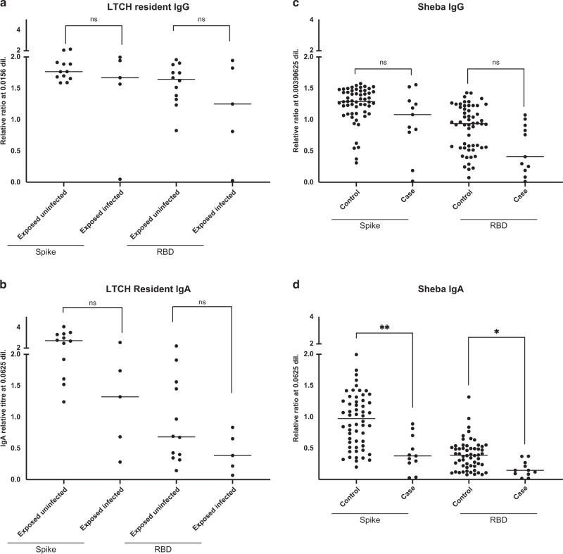
Although SARS-CoV-2 infects the upper respiratory tract, we know little about the amount, type, and kinetics of antibodies (Ab) generated in the oral cavity in response to COVID-19 vaccination. We collected serum and saliva samples from participants receiving two doses of mRNA COVID-19 vaccines and measured the level of anti-SARS-CoV-2 Ab. We detected anti-Spike and anti-Receptor Binding Domain (RBD) IgG and IgA, as well as anti-Spike/RBD associated secretory component in the saliva of most participants after dose 1. Administration of a second dose of mRNA boosted the IgG but not the IgA response, with only 30% of participants remaining positive for IgA at this timepoint. At 6 months post-dose 2, these participants exhibited diminished anti-Spike/RBD IgG levels, although secretory component-associated anti-Spike Ab were more stable. Examining two prospective cohorts we found that participants who experienced breakthrough infections with SARS-CoV-2 variants had lower levels of vaccine-induced serum anti-Spike/RBD IgA at 2-4 weeks post-dose 2 compared to participants who did not experience an infection, whereas IgG levels were comparable between groups. These data suggest that COVID-19 vaccines that elicit a durable IgA response may have utility in preventing infection. Our study finds that a local secretory component-associated IgA response is induced by COVID-19 mRNA vaccination that persists in some, but not all participants. The serum and saliva IgA response modestly correlate at 2-4 weeks post-dose 2. Of note, levels of anti-Spike serum IgA (but not IgG) at this timepoint are lower in participants who subsequently become infected with SARS-CoV-2. As new surges of SARS-CoV-2 variants arise, developing COVID-19 booster shots that provoke high levels of IgA has the potential to reduce person-to-person transmission.


Conflict of interest statement

The authors declare no competing interests.

Figures

None
Graphical abstract
Fig. 1
Fig. 1
Analysis of anti-Spike and anti-RBD antibodies in saliva from participants receiving COVID-19 mRNA vaccines. AD Anti-Spike/RBD IgG and IgA were measured in the saliva of vaccinated LTCH workers after two doses of mRNA vaccine (n = 107) and compared to COVID-19 convalescent controls (n = 11) and individually run negative controls (n = 9). The positive cutoff (dotted line) was calculated as 2 standard deviations above the mean of a pool of negative control samples (n = 51) for each individual assay. EH Anti-Spike/RBD IgG and IgA were also analyzed in the saliva of vaccinated participants that were followed at sequential timepoints before and after dose 1 and dose 2 of mRNA vaccination (MSB-1, n = 27, with n = 13 participants at 3 months post-dose 1, and n = 26 at 2 weeks post-dose 2). All data are expressed as a percentage of a pooled positive plate control of n = 11 COVID-19 convalescent saliva (see Methods). Solid black bars denote the median for each cohort. Mann–Whitney U test was used to calculate significance for (AD). For (EH) a Friedman pair-wise multiple comparison test was used for pair-wise comparisons for multiple timepoints, considering only data points from participants who had completed all timepoints (n = 11). *p < 0.05; **p < 0.01; ***p < 0.001; ****p < 0.0001. † = we were only able to recall n = 13 of the participants at 3 months post-dose 1.
Fig. 2
Fig. 2
Detection of secretory component associated with anti-Spike and anti-RBD antibodies in saliva from participants receiving COVID-19 mRNA vaccines. An ELISA-based method was used to detect secretory component associated with anti-Spike (A) and anti-RBD (B) antibodies in the saliva of 2-dose vaccinated subjects (n = 42), as well as saliva taken from COVID-19 negative and positive patients (n = 21 and 11, respectively). C, D Subjects vaccinated with 2 doses of either BNT162b2 or mRNA1273 were grouped based on anti-NP+ (n = 18) vs. anti-NP− (n = 93) antibody status, which is indicative of previous infection. E, F 2-dose vaccinated subjects were also subset into those that were considered above (n = 30) or below (n = 19) the positive cutoff for salivary IgA and analyzed for secretory component. Secretory component associated with anti-Spike (G) and anti-RBD (H) antibodies was also analyzed in samples collected post-dose 1 (n = 27) and post-dose 2 (n = 26) from MSB-1. Solid black bars denote the median for each cohort, while the dotted black line denotes the positive cutoff, calculated as 2 standard deviations above the mean of a pool of negative control samples Mann–Whitney U test was used to calculate significance, with ns not significant, **p < 0.01; ***p < 0.001; ****p < 0.0001.
Fig. 3
Fig. 3
Different decay kinetics of anti-Spike and anti-RBD IgG versus IgA in saliva from participants receiving COVID-19 mRNA vaccines. Saliva from n = 32 LTCH participants was assessed for the presence of IgG (A, B), IgA (C, D), and SIgA (E, F) antibodies against Spike (A, C, E) or RBD (B, D, F). The Wilcoxon signed-rank test was used to calculate significance between groups. ns not significant, *p < 0.05; **p < 0.01; ***p < 0.001; ****p < 0.0001.
Fig. 4
Fig. 4
Participants who experience a breakthrough infection have lower level of anti-Spike/RBD IgA at 2–4 weeks post-vaccination. Serum samples from vaccinated Toronto LTCH residents were taken at 2–4 weeks post-dose 2. Serum anti-Spike/RBD IgG (A) and IgA (B) levels were compared in participants who were subsequently exposed to P.1 gamma SARS-CoV-2 and either infected (exposed infected, n = 5) or not infected (exposed uninfected, n = 12). In a separate cohort of double vaccinated healthcare workers from the Sheba Medical Center in Ramat Gan, Israel, serum samples were taken at 2–4 weeks post-dose 2. Serum anti-Spike/RBD IgG (C) and IgA (D) levels were compared in participants who experienced a breakthrough infection (cases, n = 11) vs. controls who did not (control, n = 56). Solid black lines denote the median for each cohort. Kruskal–Wallis test with a correction for multiple comparisons was used to conduct statistical analysis between groups. NS not significant, *p < 0.5; **p < 0.01.

References

    1. Walls AC, et al. Structure, function, and antigenicity of the SARS-CoV-2 spike glycoprotein. Cell. 2020;181:281–292. doi: 10.1016/j.cell.2020.02.058. 1:CAS:528:DC%2BB3cXkvVejsLk%3D. - DOI - PMC - PubMed
    1. Zou L, et al. SARS-CoV-2 viral load in upper respiratory specimens of infected patients. N. Engl. J. Med. 2020;382:1177–1179. doi: 10.1056/NEJMc2001737. - DOI - PMC - PubMed
    1. Isho B., et al. Persistence of serum and saliva antibody responses to SARS-CoV-2 spike antigens in COVID-19 patients. Sci. Immunol. 2020;5:eabe5511. - PMC - PubMed
    1. Huang N, et al. SARS-CoV-2 infection of the oral cavity and saliva. Nat. Med. 2021;27:892–903. doi: 10.1038/s41591-021-01296-8. 1:CAS:528:DC%2BB3MXnsFKjs7o%3D. - DOI - PMC - PubMed
    1. Stadtmueller B.M., et al. The structure and dynamics of secretory component and its interactions with polymeric immunoglobulins. Elife. 2016;5:e10640. - PMC - PubMed

Publication types

Supplementary concepts

Grants and funding