. 2022 Apr 25;13(1):2225.
doi: 10.1038/s41467-022-29862-9.

An unexpected strategy to alleviate hypoxia limitation of photodynamic therapy by biotinylation of photosensitizers

Affiliations

An unexpected strategy to alleviate hypoxia limitation of photodynamic therapy by biotinylation of photosensitizers

Jing An et al. Nat Commun. .

Abstract

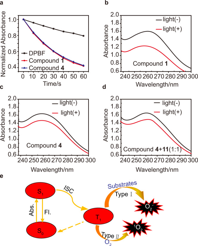
The most common working mechanism of photodynamic therapy is based on high-toxicity singlet oxygen, which is called Type II photodynamic therapy. But it is highly dependent on oxygen consumption. Recently, Type I photodynamic therapy has been found to have better hypoxia tolerance to ease this restriction. However, few strategies are available on the design of Type I photosensitizers. We herein report an unexpected strategy to alleviate the limitation of traditional photodynamic therapy by biotinylation of three photosensitizers (two fluorescein-based photosensitizers and the commercially available Protoporphyrin). The three biotiylated photosensitizers named as compound 1, 2 and 3, exhibit impressive ability in generating both superoxide anion radicals and singlet oxygen. Moreover, compound 1 can be activated upon low-power white light irradiation with stronger ability of anion radicals generation than the other two. The excellent combinational Type I / Type II photodynamic therapy performance has been demonstrated with the photosensitizers 1. This work presents a universal protocol to provide tumor-targeting ability and enhance or trigger the generation of anion radicals by biotinylation of Type II photosensitizers against tumor hypoxia.


Conflict of interest statement

The authors declare no competing interests.

Figures

Fig. 1
Fig. 1. The synthesis route for biotinylation of photosensitizers from PS-COOH to PS-Biotin.
a The carboxyl-terminal photosensitizer PS-COOH is condensed with hydrazine modified biotin to form PS-Biotin. b Chemical structures of compounds (PS-Biotin and PS-COOH) 1–6.
Fig. 2
Fig. 2. Absorption spectra and emission spectra of compounds 16.
Absorption spectra of (a) compound 1 and 4, (c) 2 and 5, (e) 3 and 6. Emission spectra of (b) 1 and 4 (λex = 640 nm), (d) 2 and 5 (λex = 469 nm), (f) 3 and 6 (λex = 403 nm) in ethanol. Fl fluorescence.
Fig. 3
Fig. 3. ROS generation of compound 1.
a Normalized absorbance of DPBF (50 μM) at 411 nm in the presence of compound 1 (10 μM) or 4 (10 μM) as a function of irradiation time (white light: 20 mW/cm2). bd Degradation of NBT (24 μM) by O2−• under different treatments (“+” represents with irradiation, “−” represents without irradiation, white light 20 mW/cm2, 3 min). e Schematic illustration of O2−• and 1O2 generation. Abs. Absorb, Fl fluorescence, ISC Intersystem Crossing, S0 ground state, S1 singlet state, T1 triplet state.
Fig. 4
Fig. 4. Cellular uptake and in vitro PDT effect of compound 1.
Subcellular colocalization images of compound 1 (10 μM) in MCF-7 cells with a lysosome-localized tracker LysoTracker Green (1 μM), b mitochondria-localized tracker Rhodamine 123 (1 μM), and c nuceu-localized tracker Hochest 33324 (1 μM), respectively. ac right panel represents the colocalization coefficient of compound 1 and each tracker is 0.83, 0.38, and 0.11, respectively. Scale bar: 30 μm. d Dark toxicity of compound 1 on COS-7 cells and MCF-7 cells under normoxia. Data in (d) are presented as mean ± s.d. derived from n = 6 independent biological samples. e Cell viability of COS-7 cells and MCF-7 cells treated with increasing concentrations of compound 1 after exposure to white-light irradiation for 10 min (white light 20 mW/cm2). Data in (e) are presented as mean ± s.d. derived from n = 5 independent biological samples. f Live/dead cell costaining assays using Calcein-AM and propidium iodide as fluorescence probes under normoxic (21% O2) or hypoxic (1% O2) conditions (green fluorescence for live cells, red fluorescence for dead cells, white light 20 mW/cm2 for 10 min). Scale bar: 100 μm.
Fig. 5
Fig. 5. In vitro ROS generation of compound 1.
CLSM images of MCF-7 cells stained with DCFH-DA (a), compound 1 (10 μM) (b), and DCFH-DA + compound 1 with different light doses under (c, d) normoxic (21% O2) and (e) hypoxic (1% O2) conditions. Ex: 488 nm, Em: 500–550 nm. Scale bar: 30 μm. CLSM images of MCF-7 cells stained with compound 1 (10 μM) under normoxic (21% O2, f) and hypoxic (1% O2, g) conditions using DHE as the intracellular O2−• fluorescence indicator before and after white-light irradiation (“+” represents with irradiation, “−” represents without irradiation, white light 20 mW/cm2, 10 min). Ex: 488 nm, Em: 590–630 nm. Scale bar: 30 μm. Each experiment was repeated three times independently, with similar results.
Fig. 6
Fig. 6. Photocytotoxicity of compound 1 to MCF-7 cells 3D MCTS.
The CLSM images of MCTS incubated with compound 1 (10 μM) for 4 h (0 d) and then exposed to white-light irradiation (40 mW/cm2) for 20 min each day (1 d, 2 d, 3 d). The experiment was repeated three times independently, with similar results. Scale bar: 400 μm.
Fig. 7
Fig. 7. The mechanistic explanation of the biotinylation effect.
Cyclic voltammograms of a compound 1, 4, b 2, 5, and c 3, 6 in DMF with 0.1 M (n-Bu)4N+PF6 as a supporting electrolyte, glassy carbon as a working electrode, Ag/AgCl as a reference electrode, Pt wire as a counter electrode, and a scan rate of 10 mVs−1. Fc/Fc+ was used as an external reference. d Photochemical reactions during Type I mechanism (3PS* represents the triple excited state of photosensitizer, PS−• represents the anion radicals of photosensitizer, 3PS* is transformed into a radical anion by accepting electrons from adjacent substrates and giving external electrons to oxygen to form O2−•). e Optimized triplet state structure, HOMO and LUMO of compound 1 (DFT, B3LYP/6-31G (d) calculated frontier orbitals relevant to the triplet state).

References

    1. Zhang J, et al. Intrinsically cancer-mitochondria-targeted thermally activated delayed fluorescence nanoparticles for two-photon-activated fluorescence imaging and photodynamic therapy. ACS Appl. Mater. Interfaces. 2019;11:41051–41061. doi: 10.1021/acsami.9b14552. - DOI - PubMed
    1. Liu P, et al. Oxygen-self-supplying and HIF-1alpha-inhibiting core-shell nanosystem for hypoxia-resistant photodynamic therapy. ACS Appl. Mater. Interfaces. 2019;11:48261–48270. doi: 10.1021/acsami.9b18112. - DOI - PubMed
    1. Phua SZF, et al. Catalase-integrated hyaluronic acid as nanocarriers for enhanced photodynamic therapy in solid tumor. ACS Nano. 2019;13:4742–4751. doi: 10.1021/acsnano.9b01087. - DOI - PubMed
    1. Feng L, et al. Near-infrared light activation of quenched liposomal Ce6 for synergistic cancer phototherapy with effective skin protection. Biomaterials. 2017;127:13–24. doi: 10.1016/j.biomaterials.2016.11.027. - DOI - PubMed
    1. Zheng DW, et al. Carbon-dot-decorated carbon nitride nanoparticles for enhanced photodynamic therapy against hypoxic tumor via water splitting. ACS Nano. 2016;10:8715–8722. doi: 10.1021/acsnano.6b04156. - DOI - PubMed

Publication types