. 2022 Jul;192(7):1001-1015.
doi: 10.1016/j.ajpath.2022.04.002. Epub 2022 Apr 22.

Angiopoietin 2 Is Associated with Vascular Necroptosis Induction in Coronavirus Disease 2019 Acute Respiratory Distress Syndrome

Affiliations

Angiopoietin 2 Is Associated with Vascular Necroptosis Induction in Coronavirus Disease 2019 Acute Respiratory Distress Syndrome

David R Price et al. Am J Pathol. 2022 Jul.

Abstract

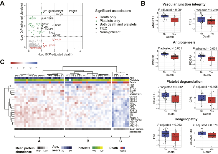
Vascular injury is a well-established, disease-modifying factor in acute respiratory distress syndrome (ARDS) pathogenesis. Recently, coronavirus disease 2019 (COVID-19)-induced injury to the vascular compartment has been linked to complement activation, microvascular thrombosis, and dysregulated immune responses. This study sought to assess whether aberrant vascular activation in this prothrombotic context was associated with the induction of necroptotic vascular cell death. To achieve this, proteomic analysis was performed on blood samples from COVID-19 subjects at distinct time points during ARDS pathogenesis (hospitalized at risk, N = 59; ARDS, N = 31; and recovery, N = 12). Assessment of circulating vascular markers in the at-risk cohort revealed a signature of low vascular protein abundance that tracked with low platelet levels and increased mortality. This signature was replicated in the ARDS cohort and correlated with increased plasma angiopoietin 2 levels. COVID-19 ARDS lung autopsy immunostaining confirmed a link between vascular injury (angiopoietin 2) and platelet-rich microthrombi (CD61) and induction of necrotic cell death [phosphorylated mixed lineage kinase domain-like (pMLKL)]. Among recovery subjects, the vascular signature identified patients with poor functional outcomes. Taken together, this vascular injury signature was associated with low platelet levels and increased mortality and can be used to identify ARDS patients most likely to benefit from vascular targeted therapies.


Figures

Figure 1
Figure 1
Study design. A: Flowchart of study subjects selected for blood proteomic profiling in the at-risk, acute respiratory distress syndrome (ARDS), and recovery cohorts. B: Venn diagram showing the overlap among subjects with COVID-19 across the study cohorts. C: Conceptual schematic of study subject sampling in relation to hospital admission (at-risk cohort), intensive care unit (ICU) admission (ARDS cohort), and ICU discharge (recovery cohort). D: Characteristics of study cohorts, including total subjects (N), blood sample type, existence of longitudinal samples, sample timing, and whether proteomic profiling, angiopoietin 2 (ANGPT2), and receptor-interacting protein kinase 3 (RIPK3) measurements were performed.
Figure 2
Figure 2
The hospitalized at-risk cohort blood proteome identifies a signature of vascular limitation preceding critical illness. A: Overview of the associations of the protein set to death and platelets. All displayed proteins were included in the final protein set. Proteins in green associated with platelets, proteins in red associated with death, and proteins in black associated with both. TIE2 was additionally included as it is the receptor for angiopoietin (ANGPT) 1 and ANGPT2. B: Box plots demonstrating the association between proteins of vascular junctional integrity, angiogenesis, platelet degranulation, and coagulopathy to mortality in the at-risk cohort after adjusting for multiple comparisons. Boxes indicate the interquartile range (IQR) of the data distribution, the line in the box represents the median value, and the whiskers extend for 1.5 times the range of the IQR. Dots indicate the protein level in individual patients. C: Heat map of protein set abundance in the at-risk COVID-19 subjects. Hierarchical clustering was performed using Ward linkage and euclidean distance. Age, platelet count, and death are overlaid at the top. Mean abundance of the 22-protein set is displayed at the bottom. Mean protein abundance is progressively lower from cluster A to B to C. N = 59 (AC).
Figure 3
Figure 3
Loss of circulating vascular proteins is associated with low platelets, mortality, and plasma angiopoietin 2 (ANGPT2) in the acute respiratory distress syndrome (ARDS) cohort. A: Heat map of 22-protein set abundance in diverse ARDS subjects, divided into two clusters. Hierarchical clustering was performed using Ward linkage and euclidean distance. Age, log10(ANGPT2), platelet count, mortality, and ARDS etiology are overlayed at the top. Mean protein abundance of the 22-protein set is overlayed at the bottom. B: Kaplan-Meier survival analysis for the two heat map clusters showing worse survival for the high ANGPT2 ARDS cluster B. The x axis was capped at 60 days. The table at the bottom indicates the number of patients at risk at each time point in the two clusters. C: Log10(ANGPT2) values in the two clusters, demonstrating higher ANGPT2 expression in low vascular protein abundance cluster B. Differential statistic was assessed with a two-sided U-test. The boxes indicate the interquartile range (IQR) of the data distribution, the line in the box represents the median value, and the whiskers extend for 1.5 times the range of the IQR. Dots indicate the protein level in individual patients across the different ARDS categories: COVID-19 (orange), bacterial sepsis (brown), and influenza (yellow). N = 60 (AC).
Figure 4
Figure 4
Angiopoietin 2 (ANGPT2) is correlated with CD61 staining microthrombi in subjects with COVID-19 acute respiratory distress syndrome (ARDS). A: Representative hematoxylin and eosin (H&E), ANGPT2, and CD61 staining in subjects with COVID-19 ARDS. H&E demonstrates alveolar septal wall thickening across displayed autopsy subjects (arrows). Increased ANGPT2 (open arrowheads) and CD61 (closed arrowheads) immunostaining is seen in subjects NA and P3 in a vascular distribution. B: Lung autopsy specimens from 20 subjects with COVID-19 ARDS were stained for ANGPT2 and platelet activation stain CD61. High ANGPT2 corresponds to autopsy subjects with ANGPT2 quantification above the median of the autopsy cohort, whereas low ANGPT2 represents the low ANGPT2 cohort. High ANGPT2 was associated with increased CD61 staining (P = 0.005). C: Blood proteomic data from autopsy subjects P1 and P2 (both low ANGPT2/low CD61 staining) and subject P3 (high ANGPT2 and high CD61) demonstrate that low expression of the vascular protein set is associated with high ANGPT2 and high CD61 staining. Angiopoietin axis proteins ANGPT1 and TIE2 highlighted in red. N = 10 for high ANGPT2 and N = 10 for low ANGPT2 (B). Scale bars = 50 μm (A). NA, no blood proteomic data available for the autopsy subject.
Figure 5
Figure 5
Induction of vascular cell death in angiopoietin 2 (ANGPT2)–associated vascular injury. A: Plasma receptor-interacting protein kinase 3 (RIPK3) in acute respiratory distress syndrome (ARDS) by heat map cluster (Figure 3A). RIPK3 is associated with high ANGPT2 (P = 0.020). COVID-19 (orange), bacterial sepsis (brown), and influenza (mustard) data are shown. B: Correlation of plasma RIPK3 and plasma ANGPT2 in the ARDS cohort (Figure 3). r indicates the Pearson correlation coefficient of the two variables, and P indicates its corresponding P value. The black line represents the linear regression line, and the gray area indicates the 95% CI of the fit. Dots indicate the protein level in individual patients across the different ARDS categories. C: Phosphorylated mixed lineage kinase domain-like (pMLKL) staining in COVID-19 ARDS autopsy subjects P1 and P2 (both low ANGPT2/low CD61 staining) and subjects NA and P3 (high ANGPT2 and high CD61), demonstrating increased expression of pMLKL (open arrowheads) in autopsy subjects with high ANGPT2 staining. N = 60 (A and B). Scale bars = 50 μm (C). NA, no blood proteomic data available for the autopsy subject.
Figure 6
Figure 6
Among subjects with COVID-19 acute respiratory distress syndrome recovery, longitudinal plasma proteomics identifies a stable protein trajectory associated with good functional recovery. A: Heat map of COVID-19 recovery subjects. Functional recovery, age, platelet count, and 12-month recovery scores are overlaid at the top. Hierarchical clustering was performed with Ward linkage and euclidean distance. B: Follow-up recovery scores at 12 months after intensive care unit (ICU) admission in the two heat map clusters. Differential statistic was assessed with a two-sided U-test. The boxes indicate the interquartile range (IQR) of the data distribution, the line in the box represents the median value, and the whiskers extend for 1.5 times the range of the IQR. Dots indicate the protein level in individual patients. High scores indicate worse functional recovery. C: Trajectory of vascular proteins from ICU to recovery time points by functional recovery group. The boxes indicate the IQR of the data distribution, the line in the box represents the median value, and the whiskers extend for 1.5 times the range of the IQR. Dots indicate the protein level in individual patients in the two time points. Values from the same patient are linked by a line and colored according to the corresponding heat map cluster: A (cream) or B (red). Differential statistic of the protein trajectories between the two patient clusters was computed with a linear model. All displayed trajectory differences were significant to an adjusted P < 0.25. N = 12 (AC).

Comment in

References

    1. Matthay M.A., Zemans R.L., Zimmerman G.A., Arabi Y.M., Beitler J.R., Mercat A., Herridge M., Randolph A.G., Calfee C.S. Acute respiratory distress syndrome. Nat Rev Dis Primers. 2019;5:18. - PMC - PubMed
    1. Meyer N.J., Calfee C.S. Novel translational approaches to the search for precision therapies for acute respiratory distress syndrome. Lancet Respir Med. 2017;5:512–523. - PMC - PubMed
    1. Agrawal A., Matthay M.A., Kangelaris K.N., Stein J., Chu J.C., Imp B.M., Cortez A., Abbott J., Liu K.D., Calfee C.S. Plasma angiopoietin-2 predicts the onset of acute lung injury in critically ill patients. Am J Respir Crit Care Med. 2013;187:736–742. - PMC - PubMed
    1. Gallagher D.C., Parikh S.M., Balonov K., Miller A., Gautam S., Talmor D., Sukhatme V.P. Circulating angiopoietin 2 correlates with mortality in a surgical population with acute lung injury/adult respiratory distress syndrome. Shock. 2008;29:656–661. - PMC - PubMed
    1. Ziegler T., Horstkotte J., Schwab C., Pfetsch V., Weinmann K., Dietzel S., Rohwedder I., Hinkel R., Gross L., Lee S., Hu J., Soehnlein O., Franz W.M., Sperandio M., Pohl U., Thomas M., Weber C., Augustin H.G., Fässler R., Deutsch U., Kupatt C. Angiopoietin 2 mediates microvascular and hemodynamic alterations in sepsis. J Clin Invest. 2013;123:3436–3445. - PMC - PubMed

Publication types