Review
. 2022 Jul;32(4):565-581.
doi: 10.1111/jon.13001. Epub 2022 Apr 25.

The tauopathies: Neuroimaging characteristics and emerging experimental therapies

Affiliations
Review

The tauopathies: Neuroimaging characteristics and emerging experimental therapies

Kalen J Riley et al. J Neuroimaging. 2022 Jul.

Abstract

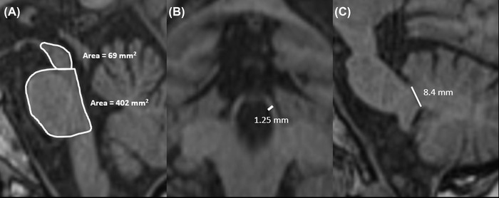
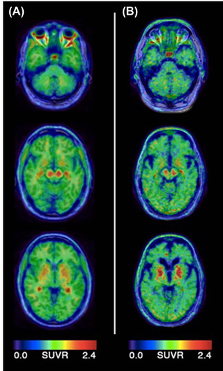
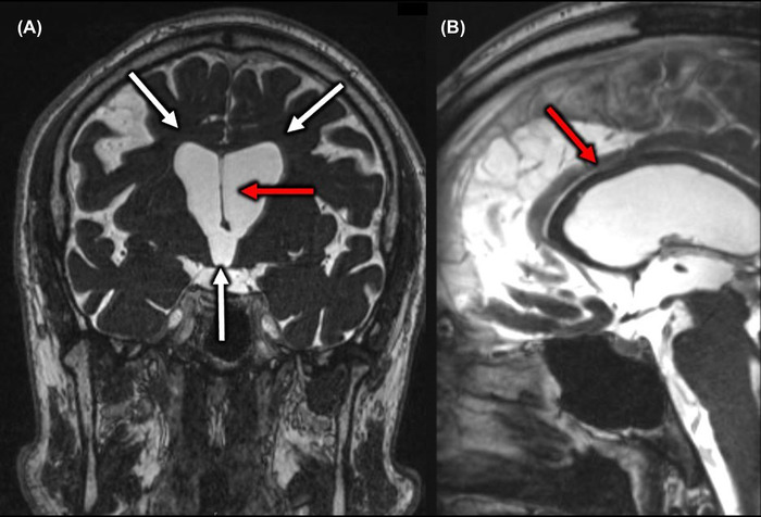
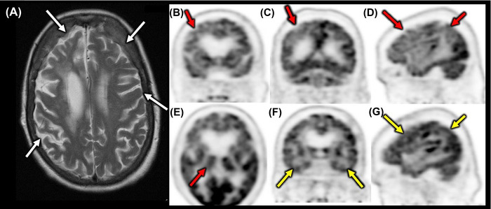
The tauopathies are a heterogeneous group of neurodegenerative disorders in which the prevailing underlying disease process is intracellular deposition of abnormal misfolded tau protein. Diseases often categorized as tauopathies include progressive supranuclear palsy, chronic traumatic encephalopathy, corticobasal degeneration, and frontotemporal lobar degeneration. Tauopathies can be classified through clinical assessment, imaging findings, histologic validation, or molecular biomarkers tied to the underlying disease mechanism. Many tauopathies vary in their clinical presentation and overlap substantially in presentation, making clinical diagnosis of a specific primary tauopathy difficult. Anatomic imaging findings are also rarely specific to a single tauopathy, and when present may not manifest until well after the point at which therapy may be most impactful. Molecular biomarkers hold the most promise for patient care and form a platform upon which emerging diagnostic and therapeutic applications could be developed. One of the most exciting developments utilizing these molecular biomarkers for assessment of tau deposition within the brain is tau-PET imaging utilizing novel ligands that specifically target tau protein. This review will discuss the background, significance, and clinical presentation of each tauopathy with additional attention to the pathologic mechanisms at the protein level. The imaging characteristics will be outlined with select examples of emerging imaging techniques. Finally, current treatment options and emerging therapies will be discussed. This is by no means a comprehensive review of the literature but is instead intended for the practicing radiologist as an overview of a rapidly evolving topic.

Keywords: chronic traumatic encephalopathy; corticobasal degeneration; frontotemporal lobar degeneration; molecular imaging; progressive supranuclear palsy; tau; tauopathies.


Conflict of interest statement

The authors declare no conflict of interest.

Figures

FIGURE 1
FIGURE 1
4 tau real‐time quaking‐induced conversion (4T RT‐QuIC) assay for tauopathy detection. The patient tissue sample with suspected abnormal tau protein is delivered into a solution containing many copies of the normal recombinant tau protein. The solution is placed on a shaker for 1 minute followed by 1 minute of incubation. If abnormal tau protein is present, the normal recombinant tau proteins will become misfolded and propagate the misfolding pattern thus causing a significant amplification. The fluorescent probe will bind only the misfolded proteins and therefore the amplified signal can be detected using a fluorimeter. Normal negative controls do not fluoresce
FIGURE 2
FIGURE 2
Abnormal tau seeding and propagation. The normal tau protein (top) converts to a misfolded tau protein and starts aggregating and altering the conformation of neighboring tau proteins. The larger oligomer unit forms fibrils that then build into disorganized aggregates. The aggregates can spread interneuronally across the synapse to begin the process of destruction in neighboring neurons. Beginning locally in a given part of the brain, the disease spreads to more distant parts of the brain to induce the neurodegenerative disease (shown on the bottom). This is shown progressing from the darker blue color in the brainstem to the lightest blue color in the more distal cortical regions of the cerebrum
FIGURE 3
FIGURE 3
Hummingbird sign, midbrain to pons diameter, and midbrain to pons area ratio in progressive supranuclear palsy (PSP). Marked midbrain atrophy is present in this patient with a hummingbird appearance of the midbrain and pons in the sagittal MRI plane (A). The midbrain to pons diameter ratio is 0.51 (9.23 mm midbrain diameter/18 mm pons diameter) in this patient with cutoff for PSP of 0.52 (A). For the midbrain to pons area ratio, a ratio of the midbrain and pons areas is determined (53 mm2/455 mm2) to be 0.117 with cutoff of 0.12 (B)
FIGURE 4
FIGURE 4
Morning glory sign. Axial T1 (A) and T2 (B) MRI show lateral concavity and atrophy of the midbrain (white arrows) in a patient with progressive supranuclear palsy
FIGURE 5
FIGURE 5
MR Parkinsonism index (MRPI). Sagittal and coronal T1 images through the midbrain and pons (A), superior cerebellar peduncle (B), and the middle cerebellar peduncle (C) in a 66‐year‐old female suspected of having progressive supranuclear palsy (PSP). The MRPI is calculated by taking the ratio of the pons (P) to midbrain (M) areas multiplied by the ratio of the middle cerebellar peduncle (MCP) width to the superior cerebellar peduncle (SCP) width [(P / M) × (MCP / SCP)]. The MRPI in this patient was 39.24. An MRPI value of more than 13.55 is abnormal and suggestive of PSP
FIGURE 6
FIGURE 6
AV1451 uptake in a patient with progressive supranuclear palsy (PSP). Patients with PSP have been observed to have increased uptake of [18F]AV‐1451 (also known as flortaucipir) in the basal ganglia, thalamus, midbrain, subthalamic nuclei, and cerebellar dentate nuclei (B) when compared to healthy control patients (A) as shown above in the figure adapted with permission from Schonhaut et al.
FIGURE 7
FIGURE 7
Former football player presented in his mid‐40s with mild early atrophy and clinical concern for chronic traumatic encephalopathy (CTE). The objective of this figure is to depict the nonspecific findings of CTE via brain MRI early on. In this patient, there is mild enlargement of the lateral (white arrows) and third ventricles with notable thinning of the septum pellucidum and corpus callosum (red arrows). There was also mild nonspecific atrophy of the mesial temporal structures. Molecular image has great potential to better diagnosis CTE in this early stage
FIGURE 8
FIGURE 8
MRI and 18F‐Fluorodeoxyglucose (18F‐FDG) PET of an elderly female with suspected corticobasal syndrome. Axial T2 image shows asymmetric right greater than left frontal and parietal atrophy with particular atrophy of the right perirolandic region (white arrows) (A). FDG‐PET demonstrated hypometabolism (red arrows) involving the right frontal and right parietal lobe on the coronal view (B & C) and right sagittal view (D). There was notable right thalamic hypometabolism compared to the left (E). Note preservation of activity (yellow arrows) within the temporal lobes (F). The normal left frontoparietal lobe FDG metabolism (yellow arrows) is shown for comparison (G)
FIGURE 9
FIGURE 9
MRI and PET showing asymmetric left sided temporal atrophy. T1 coronal (A) and T2 axial (B) sequences depict the asymmetric left temporal lobe volume loss (arrows) indicative of semantic dementia. Note the dramatic temporal lobe volume loss on the left with a knife‐like morphology to the left anterior temporal lobe. 18F‐Fluorodeoxyglucose PET in the axial (C) and coronal (D) planes shows corresponding hypometabolism in the left temporal lobe with more profound atrophy of the left temporal lobe compared to the right (arrows)
FIGURE 10
FIGURE 10
18F‐Fluorodeoxyglucose (FDG) PET of behavioral‐variant frontotemporal dementia (bvFTLD). FDG PET/CT in axial, sagittal and coronal planes showing hypometabolism in the frontal and temporal lobes (arrows) classic for bvFTLD shown in grayscale PET (upper row) and PET/CT fusion (lower row)

Similar articles

Cited by

References

    1. Josephs KA. Current understanding of neurodegenerative diseases associated with the protein tau. Mayo Clin Proc 2017;92:1291‐303. - PMC - PubMed
    1. Josephs KA, Hodges JR, Snowden JS, et al. Neuropathological background of phenotypical variability in frontotemporal dementia. Acta Neuropathol 2011;122:137‐53. - PMC - PubMed
    1. Arendt T, Stieler JT, Holzer M Tau and tauopathies. Brain Res Bull 2016;126:238‐92. - PubMed
    1. Spillantini MG, Goedert M, Crowther RA, et al. Familial multiple system tauopathy with presenile dementia: a disease with abundant neuronal and glial tau filaments. Proc Natl Acad Sci USA 1997;94:4113‐8. - PMC - PubMed
    1. Kovacs GG, Ferrer I, Grinberg LT, et al. Aging‐related tau astrogliopathy (artag): harmonized evaluation strategy. Acta Neuropathol 2016;131:87‐102. - PMC - PubMed