. 2022 May 3;11(9):e025381.
doi: 10.1161/JAHA.122.025381. Epub 2022 Apr 26.

Microcirculatory Function in Nonhypertrophic and Hypertrophic Myocardium in Patients With Aortic Valve Stenosis

Affiliations

Microcirculatory Function in Nonhypertrophic and Hypertrophic Myocardium in Patients With Aortic Valve Stenosis

Muhammad Sabbah et al. J Am Heart Assoc. .

Abstract

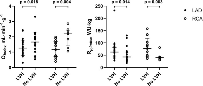
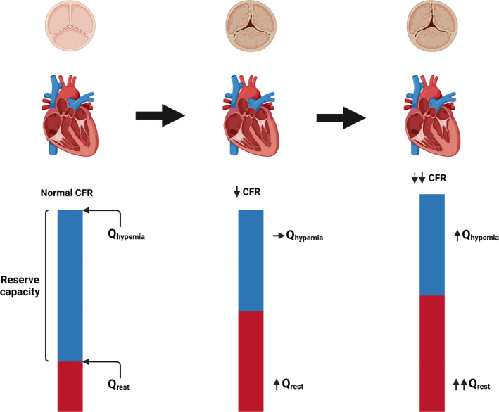
Background Left ventricular hypertrophy (LVH) has often been supposed to be associated with abnormal myocardial blood flow and resistance. The aim of this study was to evaluate and quantify the physiological and pathological changes in myocardial blood flow and microcirculatory resistance in patients with and without LVH attributable to severe aortic stenosis. Methods and Results Absolute coronary blood flow and microvascular resistance were measured using a novel technique with continuous thermodilution and infusion of saline. In addition, myocardial mass was assessed with cardiac magnetic resonance imaging. Fifty-three patients with aortic valve stenosis were enrolled in the study. In 32 patients with LVH, hyperemic blood flow per gram of tissue was significantly decreased compared with 21 patients without LVH (1.26±0.48 versus 1.66±0.65 mL·min-1·g-1; P=0.018), whereas minimal resistance indexed for left ventricular mass was significantly increased in patients with LVH (63 [47-82] versus 43 [35-63] Wood Units·kg; P=0.014). Conclusions Patients with LVH attributable to severe aortic stenosis had lower hyperemic blood flow per gram of myocardium and higher minimal myocardial resistance compared with patients without LVH.

Keywords: aortic stenosis; cardiac magnetic resonance imaging; coronary flow; left ventricular hypertrophy; microvascular function; thermodilution.


Figures

Figure 1
Figure 1. Absolute hyperemic coronary flow measurement in the LAD by continuous thermodilution.
Top traces represent aortic pressure (Pa; red) and distal coronary pressure (Pd; green), recorded simultaneously with coronary temperature (blue trace). The x axis represents time in seconds. The guidewire tip is positioned distally in the left anterior decending artery (LAD) and the side holes of the infusion catheter in the proximal LAD. Yellow arrow: before start of saline infusion, temperature is zeroed (ie, set equal to body temperature). Red arrow: infusion of room temperature saline at 20 mL/min starts and induces steady‐state hyperemia in the LAD and steady‐state decrease in temperature is observed after mixing of saline with blood (*). Green arrow: the sensor is pulled back to the tip of the infusion catheter to measure the infusion temperature, close to the tip of the infusion catheter. Blue arrow: infusion of saline is stopped, and temperature quickly returns to baseline. FFR indicates fractional flow reserve; and LAD, left anterior descending artery.
Figure 2
Figure 2. Correlation between absolute minimal microvascular resistance in the left anterior decending artery (LAD) and left ventricular mass (LVM) in patients with vs without left ventricular hypertrophy (LVH).
In patients without LVH, there was no correlation between absolute minimal microvascular resistance (R µ ,LAD) and LVM. In contrast, patients with LVH showed a clear negative correlation. Data are shown with omission of one extreme outlier in the LVH group. When included, r=−0.50 (P=0.008). LAD indicates left anterior descending artery; and WU, Wood Units.
Figure 3
Figure 3. Mass‐indexed hyperemic coronary blood flow and minimal microvascular resistance in the LAD and right coronary artery (RCA), according to left ventricular hypertrophy (LVH) status.
Left: Hyperemic coronary blood flow in the left anterior decending artery (LAD) and right coronary artery (RCA) indexed to left ventricular mass (LVM) (Q index ). Data show mean±SD for patients with LVH vs patients without LVH. Right: Minimal microvascular resistance (in Wood Units [WU], mm Hg∙min∙L−1) indexed to LVM (R µ,index ) in the LAD and RCA for patients with LVH vs no LVH. Data show median and interquartile range.
Figure 4
Figure 4. Relationship between myocardial mass, coronary blood flow, and minimal microvascular resistance in the left anterior descending artery (LAD) and right coronary artery (RCA).
Left: Correlation between hyperemic blood flow per gram of myocardium (Q index ) and left ventricular mass index (LVMi), showing decreasing hyperemic capacity with increasing severity of left ventricular hypertrophy. Data shown for the left anterior decending artery (LAD) and right coronary artery (RCA). Right: Correlation between minimal microvascular resistance (in Wood Units [WU], mm Hg∙min∙L−1) indexed to left ventricular mass (R µ,index ), showing increased resistance with increasing LVMi. Data shown for LAD and RCA.
Figure 5
Figure 5. Dynamic changes in coronary flow reserve (CFR) in severe aortic stenosis (AS).
Illustration of the continuum (left to right) going from a normal state to severe AS, and finally to severe AS with left ventricular hypertrophy. In the normal state, the difference between global resting and hyperemic flow (Qrest and Qhyperemia, respectively) ensures a normal CFR. When AS develops, resting flow increases with no change in hyperemic flow, thus leading to a decrease in CFR. When the myocardium hypertrophies, so does resting flow and CFR declines further. However, the further decline in CFR is attenuated by a concomitant increase in global hyperemic flow.

References

    1. Cioffi G, Faggiano P, Vizzardi E, Tarantini L, Cramariuc D, Gerdts E, de Simone G. Prognostic effect of inappropriately high left ventricular mass in asymptomatic severe aortic stenosis. Heart. 2011;97:301–307. doi: 10.1136/hrt.2010.192997 - DOI - PubMed
    1. Treibel TA, Kozor R, Schofield R, Benedetti G, Fontana M, Bhuva AN, Sheikh A, López B, González A, Manisty C, et al. Reverse myocardial remodeling following valve replacement in patients with aortic stenosis. J Am Coll Cardiol. 2018;71:860–871. doi: 10.1016/j.jacc.2017.12.035 - DOI - PMC - PubMed
    1. Rajappan K, Rimoldi OE, Dutka DP, Ariff B, Pennell DJ, Sheridan DJ, Camici PG. Mechanisms of coronary microcirculatory dysfunction in patients with aortic stenosis and angiographically normal coronary arteries. Circulation. 2002;105:470–476. doi: 10.1161/hc0402.102931 - DOI - PubMed
    1. Beach JM, Mihaljevic T, Rajeswaran J, Marwick T, Edwards ST, Nowicki ER, Thomas J, Svensson LG, Griffin B, Gillinov AM, et al. Ventricular hypertrophy and left atrial dilatation persist and are associated with reduced survival after valve replacement for aortic stenosis. J Thorac Cardiovasc Surg. 2014;147:362–369.e8. doi: 10.1016/j.jtcvs.2012.12.016 - DOI - PubMed
    1. Cortigiani L, Rigo F, Gherardi S, Bovenzi F, Molinaro S, Picano E, Sicari R. Coronary flow reserve during dipyridamole stress echocardiography predicts mortality. JACC Cardiovasc Imaging. 2012;5:1079–1085. doi: 10.1016/j.jcmg.2012.08.007 - DOI - PubMed

Publication types

MeSH terms

LinkOut - more resources