Review
. 2022 May;42(5):374-400.
doi: 10.1002/cac2.12294. Epub 2022 Apr 26.

Functions and clinical significance of mechanical tumor microenvironment: cancer cell sensing, mechanobiology and metastasis

Affiliations
Review

Functions and clinical significance of mechanical tumor microenvironment: cancer cell sensing, mechanobiology and metastasis

Hanying Zhou et al. Cancer Commun (Lond). 2022 May.

Abstract

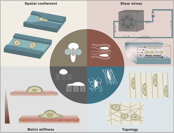
Dynamic and heterogeneous interaction between tumor cells and the surrounding microenvironment fuels the occurrence, progression, invasion, and metastasis of solid tumors. In this process, the tumor microenvironment (TME) fractures cellular and matrix architecture normality through biochemical and mechanical means, abetting tumorigenesis and treatment resistance. Tumor cells sense and respond to the strength, direction, and duration of mechanical cues in the TME by various mechanotransduction pathways. However, far less understood is the comprehensive perspective of the functions and mechanisms of mechanotransduction. Due to the great therapeutic difficulties brought by the mechanical changes in the TME, emerging studies have focused on targeting the adverse mechanical factors in the TME to attenuate disease rather than conventionally targeting tumor cells themselves, which has been proven to be a potential therapeutic approach. In this review, we discussed the origins and roles of mechanical factors in the TME, cell sensing, mechano-biological coupling and signal transduction, in vitro construction of the tumor mechanical microenvironment, applications and clinical significance in the TME.

Keywords: cytoskeleton remodeling; mechanical model; mechanosensing; mechanotransduction; tumor microenvironment.


Conflict of interest statement

The authors declare that they have no competing interests.

Figures

FIGURE 1
FIGURE 1
Properties of the TME. As a comprehensive environment, we suggest that the TME is mainly divided into two parts: (i) the biochemical microenvironment and (ii) the mechanical microenvironment. Due to the changes in biochemical components in the TME, the physical properties are dramatically changed. Uncontrolled proliferation of tumor cells constantly compresses adjacent tissues and tumor vasculature, generating internal high‐pressure solid stress and further resulting in increased blood/interstitial pressure. The imbalance between pro‐ and antiangiogenic factors causes abnormal tumor vasculature that generates much pressure on the TME: this pressure is called fluid stress. Alterations in the composition and density of the ECM occur frequently in tumors. CAFs are constantly remodeling (by depositing or cross‐linking) the ECM to cope with mechanical stress in the TME, leading to increased stiffness and altered topology. Stiffness (rigidity) refers to the resistance to deformation in response to applied force, which is measured as Young's elastic modulus. Topology (architecture) comprises ECM porosity, fibril orientation and other fibril characteristics, which wield important influences on cell polarity and function. The subtle and mutualistic relationship between tumor cells and their TME properties is formed early in solid tumor growth and evolves throughout the complete life cycle. Abbreviations: TME, tumor microenvironment; CAFs, cancer‐associated fibroblasts; ECM, extracellular matrix
FIGURE 2
FIGURE 2
In vitro models of the TME. Eager to explore the specific interactions between mechanical cues and tumorigenesis in the TME, several in vitro research models have emerged as the times require, and we mainly summarize them into four categories:(i) confinement models, (ii) shear stress models, (iii) stiffness models, and (iv) topology models. Confinement models are used to simulate in vivo ductal structures and microchannels and provide confinement paths for cell invasion and migration. When entering the vascular system, CTCs become suspended cells and remain in blood vessels, where they experience considerable fluid shear stress. Considered to be the most common and recognized mechanical abnormality of the TME, increased tissue stiffness has widely been used as a diagnostic marker and prognostic factor. Specific topology formed by component changes and special arrangements in the TME are involved in cell morphogenesis, cell polarity and cell function. Abbreviations: TME, tumor microenvironment; CTCs, circulating tumor cells
FIGURE 3
FIGURE 3
Polarized migrating tumor cells and related mechanotransduction pathways. As an iterative process, mechanotransduction touches upon multiple rounds of mechanosensing, transduction and response. Force‐induced activation of mechanosensors, such as integrins, FAs and caveolin‐1, transmits the force in the TME to intracellular locations by the anchored cytoskeleton (abundant studies have confirmed that it is directly linked to many mechanosensors) or other signaling pathways, such as integrin‐FAK, Rho, and Hippo signaling. Physically connected with the internal cytoskeleton, the nucleus responds to mechanical factors in the TME throughLINC complexes and lamins, often in the form of changes in chromosomal reorganization and gene expression, thus promoting tumor cell migration. Abbreviations: FAs, focal adhesions; TME, tumor microenvironment; LINC, linker of nucleoskeleton and cytoskeleton; NPC, nuclear pore complex; MTOC, microtubule organizing center; TKR, tyrosine kinase receptor; FAK, focal adhesion kinase; GPCRs, G‐coupled receptors; ER, endoplasmic reticulum

References

    1. Ohlund D, Elyada E, Tuveson D. Fibroblast heterogeneity in the cancer wound. J Exp Med. 2014;211(8):1503–23. - PMC - PubMed
    1. Ponten F, Ren Z, Nister M, Westermark B, Ponten J. Epithelial‐stromal interactions in basal cell cancer: the PDGF system. J Invest Dermatol. 1994;102(3):304–9. - PubMed
    1. Haak AJ, Tan Q, Tschumperlin DJ. Matrix biomechanics and dynamics in pulmonary fibrosis. Matrix Biol. 2018;73:64–76. - PMC - PubMed
    1. Santos A, Lagares D. Matrix Stiffness: the Conductor of Organ Fibrosis. Curr Rheumatol Rep. 2018;20(1):2. - PubMed
    1. Morikawa S, Baluk P, Kaidoh T, Haskell A, Jain RK, McDonald DM. Abnormalities in pericytes on blood vessels and endothelial sprouts in tumors. Am J Pathol. 2002;160(3):985–1000. - PMC - PubMed

Publication types