Multicenter Study
. 2022 Aug;304(2):419-428.
doi: 10.1148/radiol.210385. Epub 2022 Apr 26.

Quantitative MRI Characterization of the Extremely Preterm Brain at Adolescence: Atypical versus Neurotypical Developmental Pathways

Collaborators, Affiliations
Multicenter Study

Quantitative MRI Characterization of the Extremely Preterm Brain at Adolescence: Atypical versus Neurotypical Developmental Pathways

Ryan McNaughton et al. Radiology. 2022 Aug.

Abstract

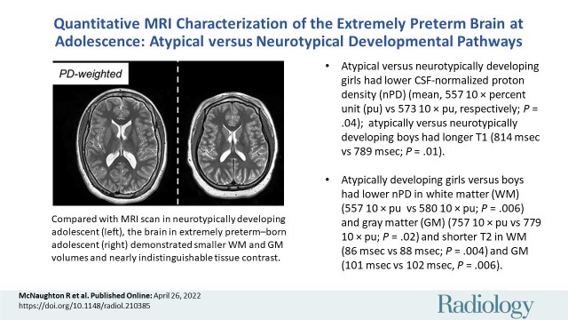
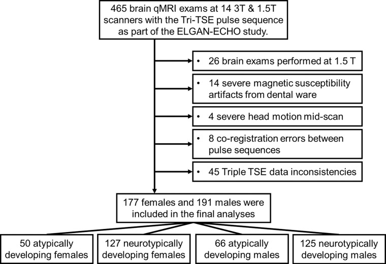
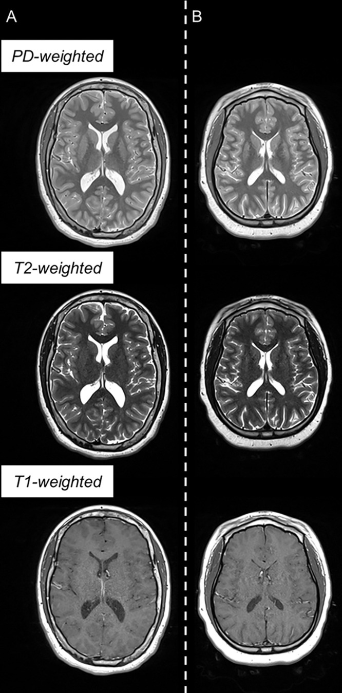
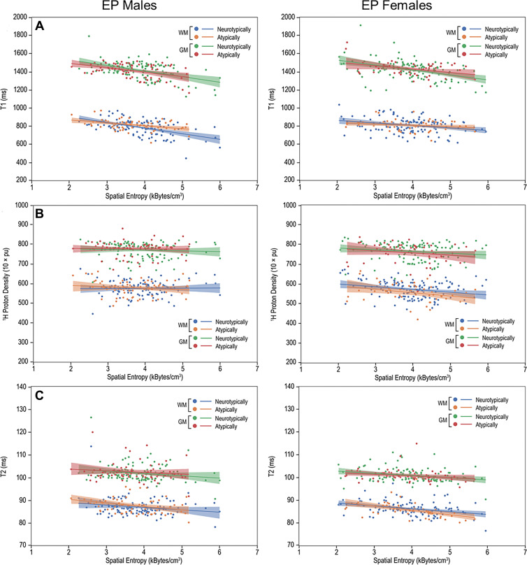
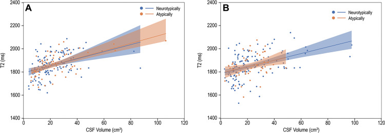
Background Extremely preterm (EP) birth is associated with higher risks of perinatal white matter (WM) injury, potentially causing abnormal neurologic and neurocognitive outcomes. MRI biomarkers distinguishing individuals with and without neurologic disorder guide research on EP birth antecedents, clinical correlates, and prognoses. Purpose To compare multiparametric quantitative MRI (qMRI) parameters of EP-born adolescents with autism spectrum disorder, cerebral palsy, epilepsy, or cognitive impairment (ie, atypically developing) with those without (ie, neurotypically developing), characterizing sex-stratified brain development. Materials and Methods This prospective multicenter study included individuals aged 14-16 years born EP (Extremely Low Gestational Age Newborns-Environmental Influences on Child Health Outcomes Study, or ELGAN-ECHO). Participants underwent 3.0-T MRI evaluation from 2017 to 2019. qMRI outcomes were compared for atypically versus neurotypically developing adolescents and for girls versus boys. Sex-stratified multiple regression models were used to examine associations between spatial entropy density (SEd) and T1, T2, and cerebrospinal fluid (CSF)-normalized proton density (nPD), and between CSF volume and T2. Interaction terms modeled differences in slopes between atypically versus neurotypically developing adolescents. Results A total of 368 adolescents were classified as 116 atypically (66 boys) and 252 neurotypically developing (125 boys) participants. Atypically versus neurotypically developing girls had lower nPD (mean, 557 10 × percent unit [pu] ± 46 [SD] vs 573 10 × pu ± 43; P = .04), while atypically versus neurotypically developing boys had longer T1 (814 msec ± 57 vs 789 msec ± 82; P = .01). Atypically developing girls versus boys had lower nPD and shorter T2 (eg, in WM, 557 10 × pu ± 46 vs 580 10 × pu ± 39 for nPD [P = .006] and 86 msec ± 3 vs 88 msec ± 4 for T2 [P = .003]). Atypically versus neurotypically developing boys had a more moderate negative association between T1 and SEd (slope, -32.0 msec per kB/cm3 [95% CI: -49.8, -14.2] vs -62.3 msec per kB/cm3 [95% CI: -79.7, -45.0]; P = .03). Conclusion Atypically developing participants showed sexual dimorphisms in the cerebrospinal fluid-normalized proton density (nPD) and T2 of both white matter (WM) and gray matter. Atypically versus neurotypically developing girls had lower WM nPD, while atypically versus neurotypically developing boys had longer WM T1 and more moderate T1 associations with microstructural organization in WM. © RSNA, 2022 Online supplemental material is available for this article.


Conflict of interest statement

Disclosures of conflicts of interest: R.M. Patent pending MR fibrography assigned to Boston University and/or Boston Medical Center. C.P. No relevant relationships. O.S. Royalties from Gakken Medical Shujunsha and Medical Sciences International; consulting fees from Boston Imaging Core Lab; honorarium from Bayer Yakuhin. J.V.R. No relevant relationships. X.Z. No relevant relationships. D.N.K. Grants from the National Institutes of Health (R01 MH083320, P41 EB019936, R24EB029173, and R25DA051675); treasurer elect of the Organization for Human Brain Mapping. J.A.F. Grants to institution from Autism Speaks, National Institute of Mental Health, Eagles Foundation, Healix, National Library of Medicine, Riccio Fund, Fulcrum, and Roche; honorarium for grand rounds from the University of Michigan. L.D. No relevant relationships. T.H. No relevant relationships. R.C.F. No relevant relationships. T.M.O. No relevant relationships. K.K.K. No relevant relationships. H.J. Book royalties from World Scientific Publishing; patents or patents pending on qMRI techniques and MR fibrography assigned to Boston University and/or Boston Medical Center; member of the Radiology Editorial Board.

Figures

None
Graphical abstract
Flowchart of participant selection from each of the 12 participating
institutions. ELGAN-ECHO = Extremely Low Gestational Age
Newborns–Environmental Influences on Child Health Outcomes, qMRI =
quantitative MRI, TSE = turbo spin echo.
Figure 1:
Flowchart of participant selection from each of the 12 participating institutions. ELGAN-ECHO = Extremely Low Gestational Age Newborns–Environmental Influences on Child Health Outcomes, qMRI = quantitative MRI, TSE = turbo spin echo.
Example triple turbo spin-echo brain MRI scans in (A) neurotypically
developing and (B) atypically developing extremely preterm–born
adolescents. Proton density (PD)–weighted, T2-weighted, and
T1-weighted images are provided. MRI scans in the atypically developing
participant (full-scale intelligence quotient <85) demonstrated
qualitatively smaller white and gray matter volumes and nearly
indistinguishable tissue contrast compared with the neurotypically
developing participant.
Figure 2:
Example triple turbo spin-echo brain MRI scans in (A) neurotypically developing and (B) atypically developing extremely preterm–born adolescents. Proton density (PD)–weighted, T2-weighted, and T1-weighted images are provided. MRI scans in the atypically developing participant (full-scale intelligence quotient <85) demonstrated qualitatively smaller white and gray matter volumes and nearly indistinguishable tissue contrast compared with the neurotypically developing participant.
Multiparametric quantitative MRI parameters versus spatial entropy
density (SEd). Scatterplots and linear fitting of the mean (A) T1, (B)
normalized proton density, and (C) T2 values versus SEd of the white matter
(WM) and gray matter (GM) for neurotypically and atypically developing
extremely preterm (EP)–born adolescents. Differences in the
associations of T1 and SEd were observed in the WM between atypically and
neurotypically developing boys. pu = percent unit.
Figure 3:
Multiparametric quantitative MRI parameters versus spatial entropy density (SEd). Scatterplots and linear fitting of the mean (A) T1, (B) normalized proton density, and (C) T2 values versus SEd of the white matter (WM) and gray matter (GM) for neurotypically and atypically developing extremely preterm (EP)–born adolescents. Differences in the associations of T1 and SEd were observed in the WM between atypically and neurotypically developing boys. pu = percent unit.
T2 versus cerebrospinal fluid (CSF) volume. Scatterplots and linear
fitting of ventricular CSF relaxation times and volume for (A) boys and (B)
girls. The presence of functional abnormality had limited impact on the
association between the T2 relaxation and volume of CSF.
Figure 4:
T2 versus cerebrospinal fluid (CSF) volume. Scatterplots and linear fitting of ventricular CSF relaxation times and volume for (A) boys and (B) girls. The presence of functional abnormality had limited impact on the association between the T2 relaxation and volume of CSF.

References

    1. Whitfield JM , Peters BA , Shoemaker C . Conference summary: a celebration of a century of neonatal care . Proc Bayl Univ Med Cent 2004. ; 17 ( 3 ): 255– 258 . - PMC - PubMed
    1. Douglass LM , Heeren TC , Stafstrom CE , et al. . Cumulative incidence of seizures and epilepsy in ten-year-old children born before 28 weeks’ gestation . Pediatr Neurol 2017. ; 73 : 13– 19 . - PMC - PubMed
    1. Volpe JJ . Dysmaturation of premature brain: importance, cellular mechanisms, and potential interventions . Pediatr Neurol 2019. ; 95 : 42– 66 . - PubMed
    1. Back SA . White matter injury in the preterm infant: pathology and mechanisms . Acta Neuropathol (Berl) 2017. ; 134 ( 3 ): 331– 349 . - PMC - PubMed
    1. Lebel C , Treit S , Beaulieu C . A review of diffusion MRI of typical white matter development from early childhood to young adulthood . NMR Biomed 2019. ; 32 ( 4 ): e3778. - PubMed

Publication types