. 2022 Jul;67(1):99-111.
doi: 10.1165/rcmb.2021-0337OC.

Molecular Dynamics and Theratyping in Airway and Gut Organoids Reveal R352Q-CFTR Conductance Defect

Affiliations

Molecular Dynamics and Theratyping in Airway and Gut Organoids Reveal R352Q-CFTR Conductance Defect

Sharon L Wong et al. Am J Respir Cell Mol Biol. 2022 Jul.

Abstract

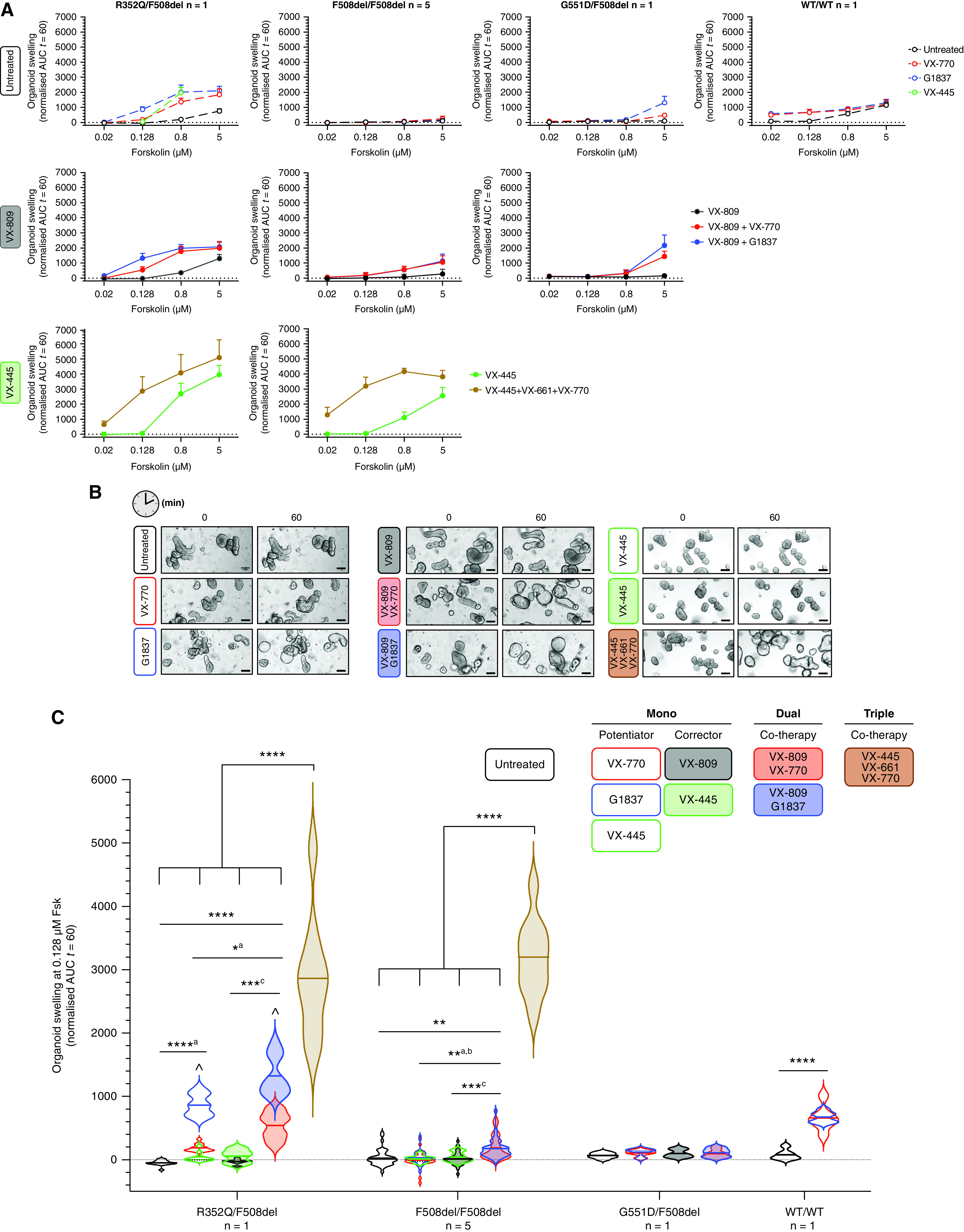
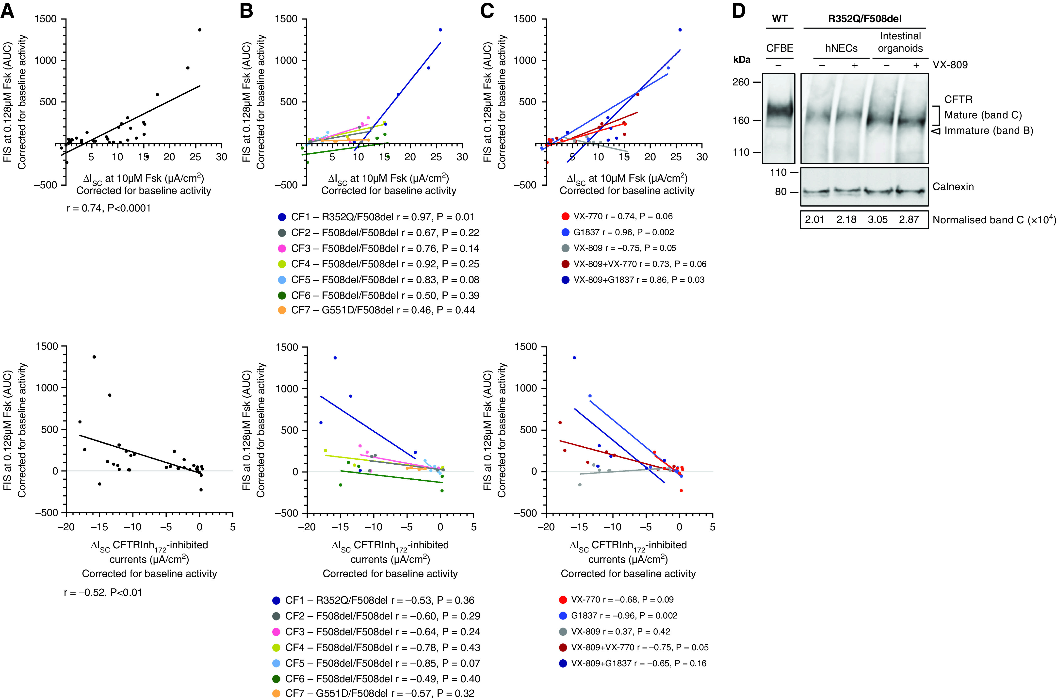
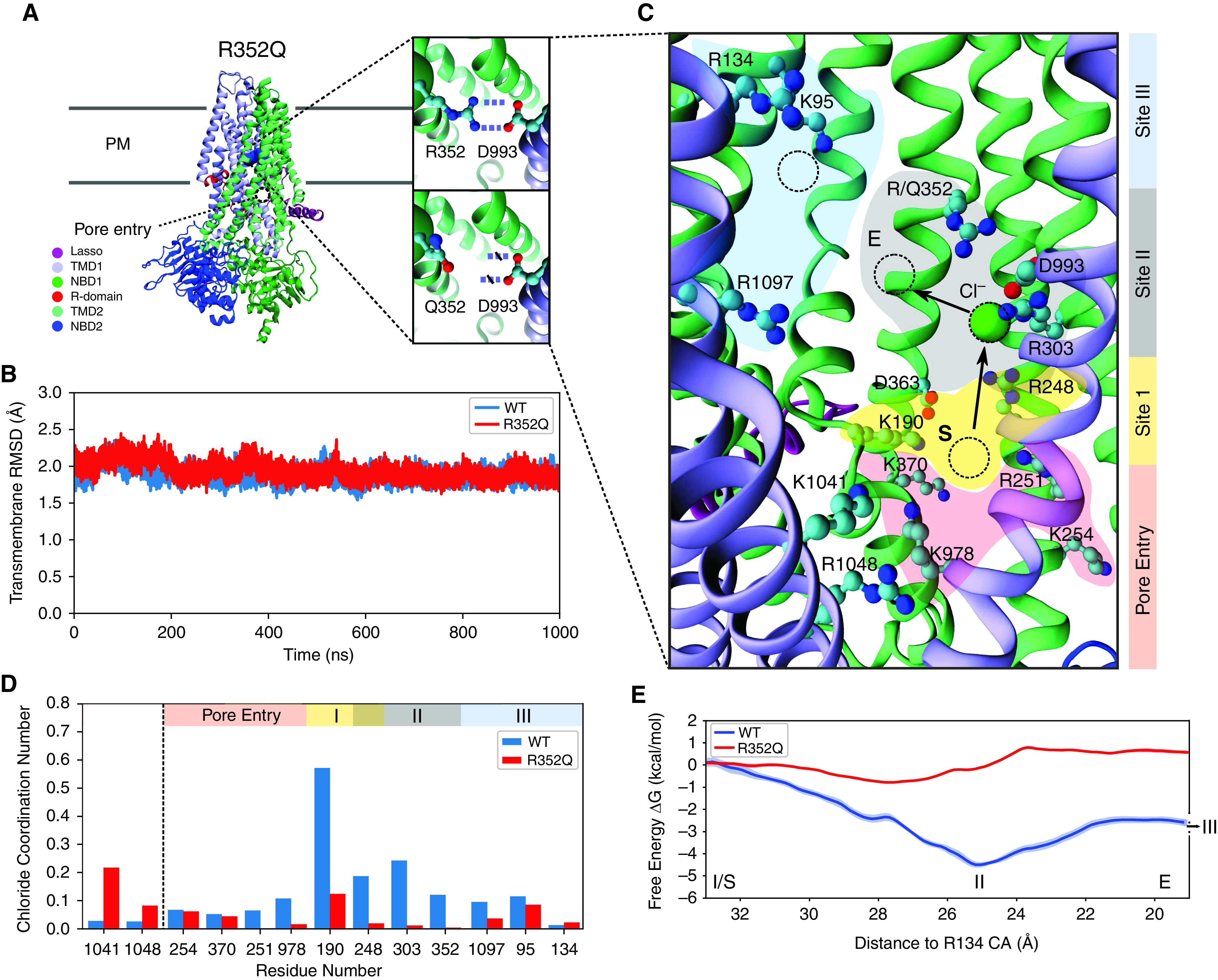
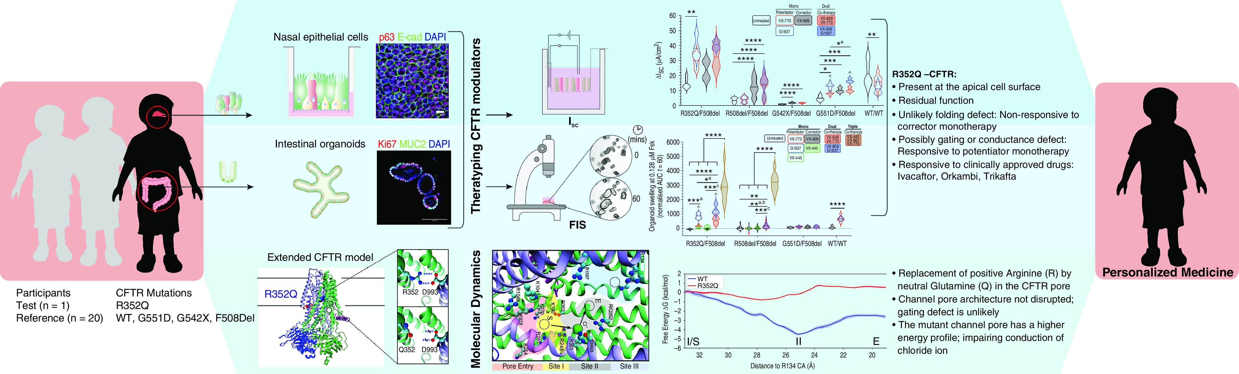
A significant challenge to making targeted cystic fibrosis transmembrane conductance regulator (CFTR) modulator therapies accessible to all individuals with cystic fibrosis (CF) are many mutations in the CFTR gene that can cause CF, most of which remain uncharacterized. Here, we characterized the structural and functional defects of the rare CFTR mutation R352Q, with a potential role contributing to intrapore chloride ion permeation, in patient-derived cell models of the airway and gut. CFTR function in differentiated nasal epithelial cultures and matched intestinal organoids was assessed using an ion transport assay and forskolin-induced swelling assay, respectively. CFTR potentiators (VX-770, GLPG1837, and VX-445) and correctors (VX-809, VX-445, with or without VX-661) were tested. Data from R352Q-CFTR were compared with data of 20 participants with mutations with known impact on CFTR function. R352Q-CFTR has residual CFTR function that was restored to functional CFTR activity by CFTR potentiators but not the corrector. Molecular dynamics simulations of R352Q-CFTR were carried out, which indicated the presence of a chloride conductance defect, with little evidence supporting a gating defect. The combination approach of in vitro patient-derived cell models and in silico molecular dynamics simulations to characterize rare CFTR mutations can improve the specificity and sensitivity of modulator response predictions and aid in their translational use for CF precision medicine.

Keywords: intestinal organoid; molecular dynamics simulations; nasal epithelial cells; personalized medicine; rare cystic fibrosis.


Figures

Figure 1.
Figure 1.
Functional response of R352Q-CFTR to cystic fibrosis transmembrane conductance regulator (CFTR) modulators in differentiated human nasal epithelial cells (hNECs). (A) Immunofluorescence staining of acetylated tubulin (ciliated cells), MUC5AC (goblet cells), and p63 (basal progenitor cells) in hNECs derived from a R352Q/F508del participant (CF1). E-cadherin (adherens junction) and ZO-1 (tight junction) are localized to the intercellular junctions of epithelial cells. The top panels are top views and the bottom panels are side views showing the pseudostratified epithelium. A 63×/1.4 numerical aperture oil immersion objective was used. Scale bars, 10 μm. (B) Dot plots of mean ciliary beat frequency (CBF) measurements (Hz) of the fully differentiated mature hNECs from participants with cystic fibrosis (CF) and without CF (wild type [WT]). Each participant is coded with a different color. Each dot represents a single field of view of CBF measurement. (C) Representative Ussing chamber recordings of short circuit current (Isc) in hNECs from participants with CF and WT-CFTR control participants. The protocol used to measure functional CFTR expression in hNECs in 0.01% DMSO vehicle (untreated; top) or pretreated with corrector (3 μM VX-809 for 48 h; bottom) followed by sequential addition of 100 μM apical amiloride (1. Amil), apical addition of either vehicle control 0.01% DMSO (black) or 10 μM VX-770 (red) or 10 μM G1837 (blue) (2. DMSO, VX-770, G1837), 10 μM basal forskolin (3. Fsk), 30 μM apical CFTR inhibitor (4. CFTRinh-172), and 100 μM apical ATP (5. ATP). A basolateral-to-apical chloride gradient was used. (D) Violin plots of total currents stimulated by DMSO or VX-770 or G1837 plus Fsk in hNECs untreated or pretreated with VX-809. Data are from 1 R352Q/F508del participant, 5 F508del/F508del participants, 3 G542X/F508del participants, and 1 G551D/F508del participant with CF and 11 WT-CFTR control participants. n, number of participants. Data are represented as violin plots to show the distribution of data. One-way ANOVA was used to determine statistically significant differences. *P < 0.05, **P < 0.01, ***P < 0.001, and ****P < 0.0001. aP for G1837 only. ^P for G1837 vs VX-770. Where VX-770 and G1837 (overlapping violin plots) both achieved statistical significance, the least significant P value is shown.
Figure 2.
Figure 2.
Functional response of R352Q-CFTR to CFTR modulators in intestinal organoids. Forskolin-induced swelling (FIS) assay in organoids (left to right) from one R352Q/F508del participant, five F508del/F508del participants, and one G551D/F508del participant with CF and one WT-CFTR control participant. Organoids were incubated overnight with 0.03% DMSO (vehicle) or 3 μM VX-809 or 3 μM VX-445 or 3 μM VX-445 + 18 μM VX-661. After 24 hours, a range of Fsk concentrations from 0.02 to 5 μM were acutely added, either alone or in combination with 3 μM VX-770 (red) or 3 μM G1837 (blue) or 3 μM VX-445 (green). (A) Values plotted are the mean ± SD of the area under the curve (AUC). (B) Representative brightfield images of R352Q/F508del organoids at baseline (t = 0) and after 1 hour of stimulation (t = 60) at 0.128 μM Fsk. Scale bars, 100 μm. (C) Violin plots of FIS response (AUC at 0.128 μM Fsk) of organoids from participants with CF and WT-CFTR control participants, expressed as the absolute AUC calculated from the time periods t = 0 (baseline) to t = 60. Data are represented as violin plots to show the distribution of data. n, number of participants. One-way ANOVA was used to determine statistically significant differences. *P < 0.05, **P < 0.01, ***P < 0.001, and ****P < 0.0001. aP for G1837 only, bP for VX-770 only, cP for VX-809 only, and ^P for G1837 versus VX-770 or VX-445. Where VX-770, G1837, and VX-445 (overlapping violin plots) achieved statistical significance, the least significant P value is shown.
Figure 3.
Figure 3.
Correlation between individual in vitro treatment-modulated CFTR function in participant-matched differentiated hNECs and intestinal organoids. Modulator-enhanced CFTR response (top) and CFTRinh-172–inhibited response (bottom), corrected for the baseline CFTR activity, in hNECs (ΔIsc) compared with organoids (FIS) from each participant shown (A) as a collective, (B) by participant, and (C) by modulator therapy. The lines through data points show simple linear regression analysis. Correlation (r) values were calculated using Pearson linear correlation. (D) Western blot in lysates from R352Q/F508del hNECs and intestinal organoids following 48-hour treatment with DMSO (vehicle) or VX-809. Cell lysates of WT-CFTR human CF bronchial epithelial (CFBE41o−) cell line were used as a control for CFTR bands B and C protein size. Densitometry was calculated by measuring the level of mature mutant CFTR (band C) normalized to the calnexin loading control. Band C represents the mature, complex-glycosylated CFTR. Band B represents the immature, core-glycosylated CFTR.
Figure 4.
Figure 4.
Simulations provide a mechanistic understanding of R352Q-CFTR dysfunction. (A) Atomistic structure of the CFTR channel pore based on cryo-EM (6MSM), with the configuration of the R352-D993 salt bridge and the disrupted R352Q highlighted in insets. Mutation of the charged R352 side chain (right top) to the neutral Q352 side chain (right bottom) disrupts the R352-D993 salt bridge present in WT-CFTR. (B) The root-mean-squared deviations (RMSDs) of the transmembrane domains of WT-CFTR (blue) and R352Q-CFTR (red) channel pore, computed using the 6MSM coordinates as reference. (C) A visualization of charged residues that line the CFTR channel pore. The path of chloride ion conduction from pore entry (pink) to site I (yellow), site II (gray), and site III (blue) is indicated. The computed free energy path in (E) is defined with reference to the chloride ion’s distance to the α-carbon of R134 in site III, with the start and end points delineated by S and E, respectively. Charged residues lining the channel pore are shown as ball and sticks. An example chloride ion is represented with a green ball in site II. (D) The histogram shows the chloride ion occupancy in WT-CFTR (blue) and R352Q-CFTR (red). The number of chloride ions within 4 Å of the sidechain nitrogen atoms of the residue in each frame of the trajectory was counted and divided by the total number of frames. (E) Free energy profile of a chloride ion in the WT-CFTR (blue) and R352Q-CFTR (red) channel pore. The change in free energy relative to site I is plotted versus the chloride ions distance to the α-carbon of R134 (site III). NBD, nucleotide-binding domain; PM, plasma membrane; R-domain, regulatory domain; TMD, transmembrane domain.
Figure 5.
Figure 5.
Flowchart demonstrating functional and in silico experiments for characterization of the R352Q-CFTR and summary of major findings.

References

    1. Ruseckaite R, Ahern S, Ranger T, Dean J, Gardam M, Bell S, et al. Australian Cystic Fibrosis Data Registry . The Australian Cystic Fibrosis Data Registry Annual Report 2017. Melbourne, Australia: Monash University, Department of Epidemiology and Preventive Medicine; March 2019. Report No. 20 [accessed 2021 Jun 30]. Available from: https://www.cysticfibrosis.org.au/getmedia/24e94d66-29fa-4e3f-8e65-21ee2...
    1. Saint-Criq V, Gray MA. Role of CFTR in epithelial physiology. Cell Mol Life Sci . 2017;74:93–115. - PMC - PubMed
    1. Marson FAL, Bertuzzo CS, Ribeiro JD. Classification of CFTR mutation classes. Lancet Respir Med . 2016;4:e37–e38. - PubMed
    1. Clancy JP, Cotton CU, Donaldson SH, Solomon GM, VanDevanter DR, Boyle MP, et al. CFTR modulator theratyping: current status, gaps and future directions. J Cyst Fibros . 2019;18:22–34. - PMC - PubMed
    1. Loo TW, Bartlett MC, Clarke DM. Corrector VX-809 stabilizes the first transmembrane domain of CFTR. Biochem Pharmacol . 2013;86:612–619. - PubMed

Publication types

MeSH terms