. 2022 Aug;92(2):304-321.
doi: 10.1002/ana.26381. Epub 2022 May 28.

Biallelic Variants in the Ectonucleotidase ENTPD1 Cause a Complex Neurodevelopmental Disorder with Intellectual Disability, Distinct White Matter Abnormalities, and Spastic Paraplegia

Daniel G Calame #  1   2   3 Isabella Herman #  1   2   3 Reza Maroofian  4 Aren E Marshall  5 Karina Carvalho Donis  6   7 Jawid M Fatih  2 Tadahiro Mitani  2 Haowei Du  2 Christopher M Grochowski  2 Sergio B Sousa  8   9 Charul Gijavanekar  2 Somayeh Bakhtiari  10   11 Yoko A Ito  5 Clarissa Rocca  4 Jill V Hunter  3   12 V Reid Sutton  2   3 Lisa T Emrick  1   2   3 Kym M Boycott  5 Alexander Lossos  13 Yakov Fellig  14 Eugenia Prus  15 Yosef Kalish  15 Vardiella Meiner  16 Manon Suerink  17 Claudia Ruivenkamp  17 Kayla Muirhead  18 Nebal W Saadi  19 Maha S Zaki  20 Arjan Bouman  21 Tahsin Stefan Barakat  21 David L Skidmore  22 Matthew Osmond  5 Thiago Oliveira Silva  7   23 David Murphy  24 Ehsan Ghayoor Karimiani  25 Yalda Jamshidi  25 Asaad Ghanim Jaddoa  26 Homa Tajsharghi  27 Sheng Chih Jin  28 Mohammad Reza Abbaszadegan  29   30 Reza Ebrahimzadeh-Vesal  30 Susan Hosseini  30 Shahryar Alavi  31 Amir Bahreini  32 Elahe Zarean  33 Mohammad Mehdi Salehi  34 Nouriya Abbas Al-Sannaa  35 Giovanni Zifarelli  36 Peter Bauer  36 Simon C Robson  37 Zeynep Coban-Akdemir  2   38 Lorena Travaglini  39   40 Francesco Nicita  39   40 Shalini N Jhangiani  41 Richard A Gibbs  41 Jennifer E Posey  2 Michael C Kruer  10   11 Kristin D Kernohan  5   42 Jonas A Morales Saute  7   43   44 Henry Houlden  4 Adeline Vanderver  18   45 Sarah H Elsea  2 Davut Pehlivan  1   2   3 Dana Marafi  2   46 James R Lupski  2   3   41   47
Affiliations

Biallelic Variants in the Ectonucleotidase ENTPD1 Cause a Complex Neurodevelopmental Disorder with Intellectual Disability, Distinct White Matter Abnormalities, and Spastic Paraplegia

Daniel G Calame et al. Ann Neurol. 2022 Aug.

Abstract

Objective: Human genomics established that pathogenic variation in diverse genes can underlie a single disorder. For example, hereditary spastic paraplegia is associated with >80 genes, with frequently only few affected individuals described for each gene. Herein, we characterize a large cohort of individuals with biallelic variation in ENTPD1, a gene previously linked to spastic paraplegia 64 (Mendelian Inheritance in Man # 615683).

Methods: Individuals with biallelic ENTPD1 variants were recruited worldwide. Deep phenotyping and molecular characterization were performed.

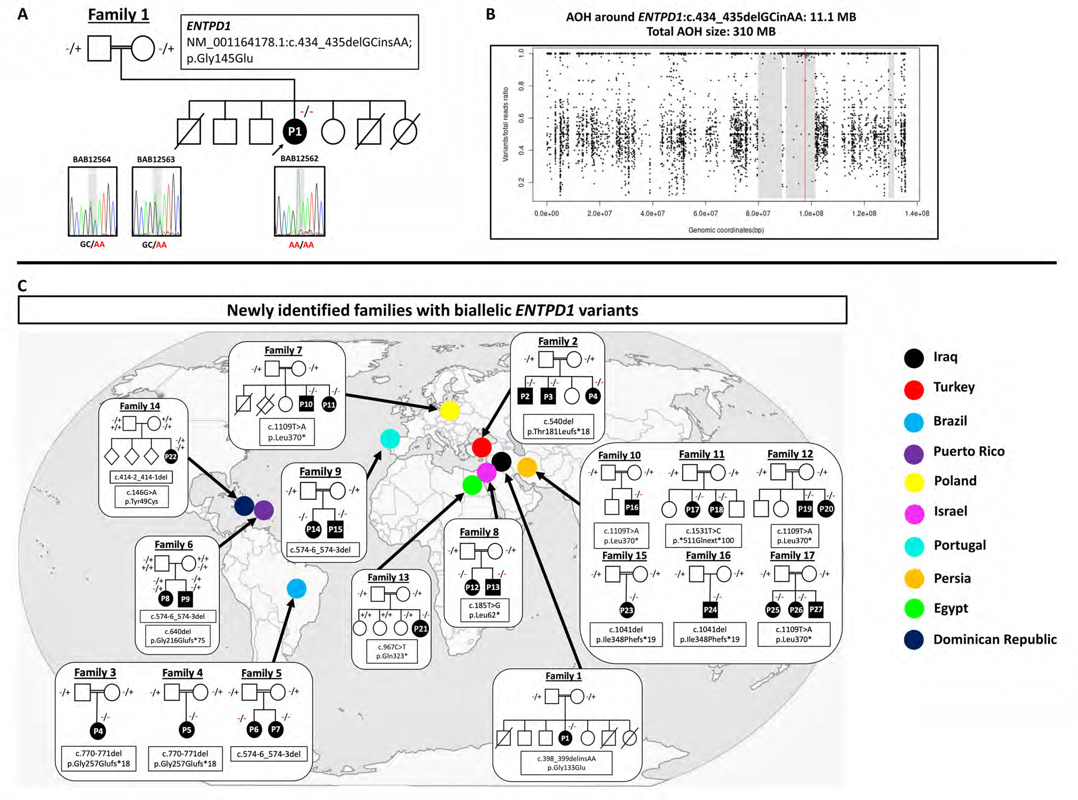
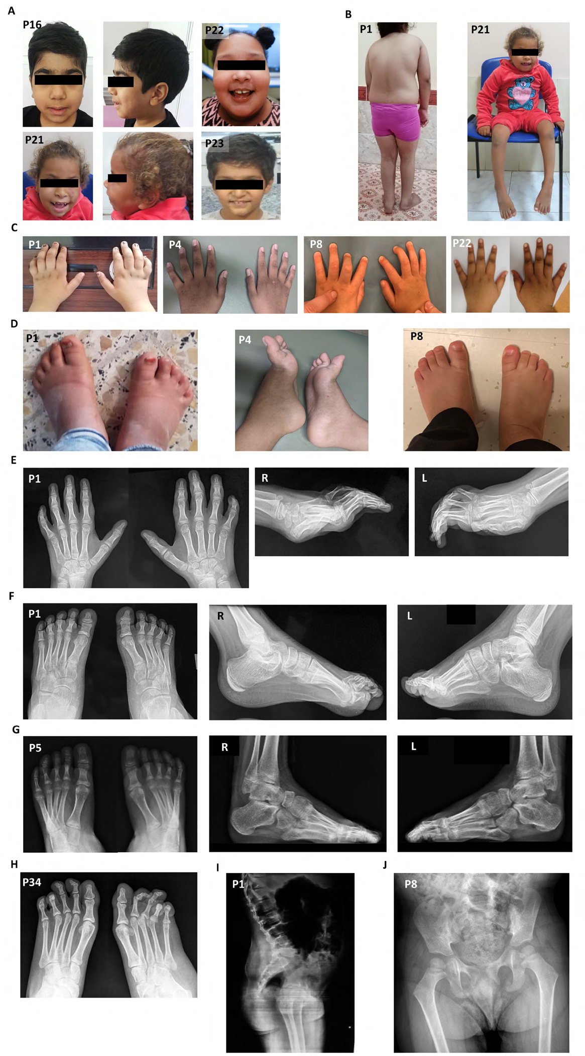
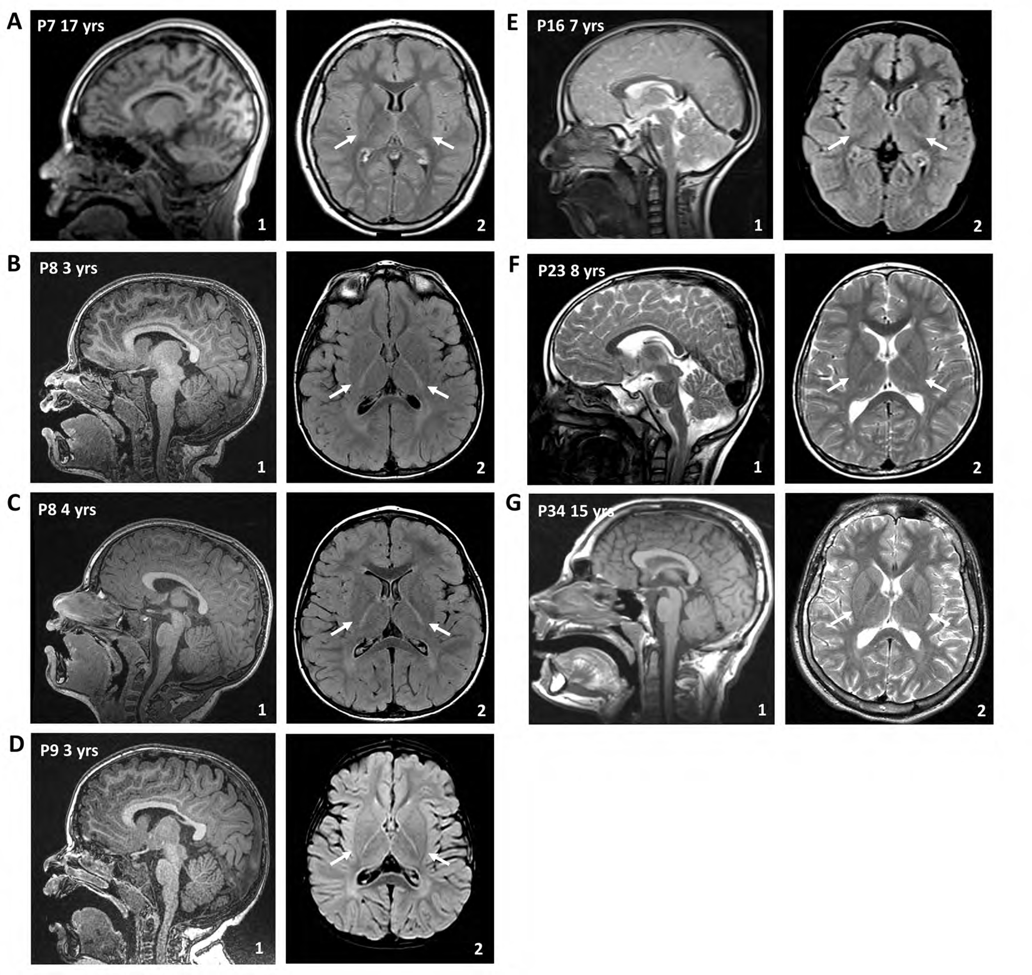
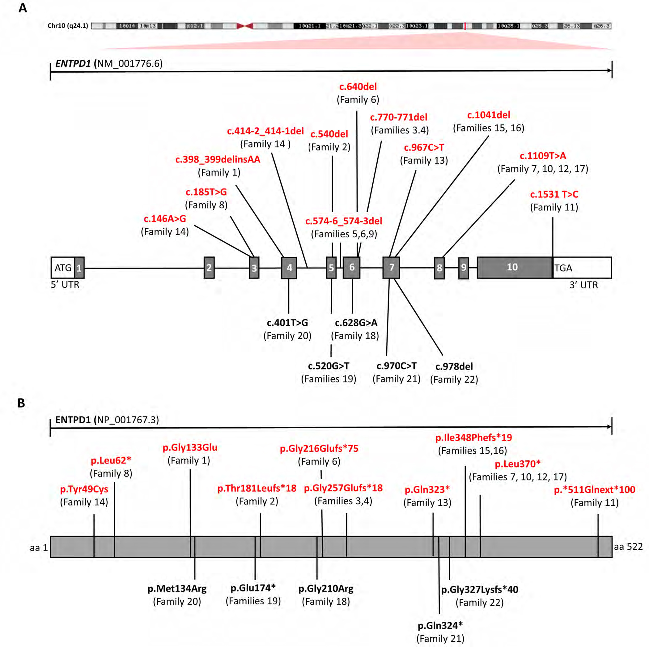
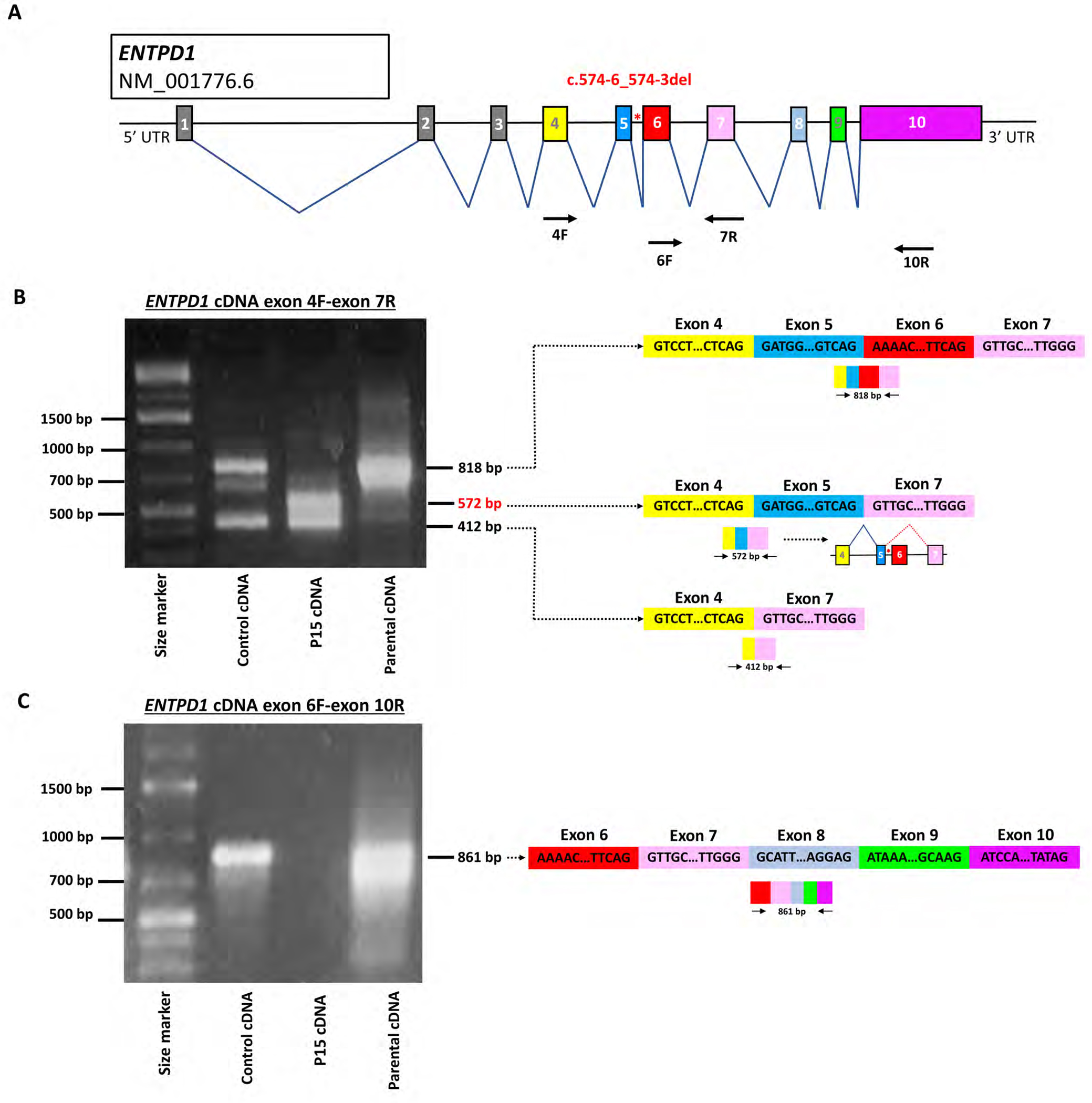
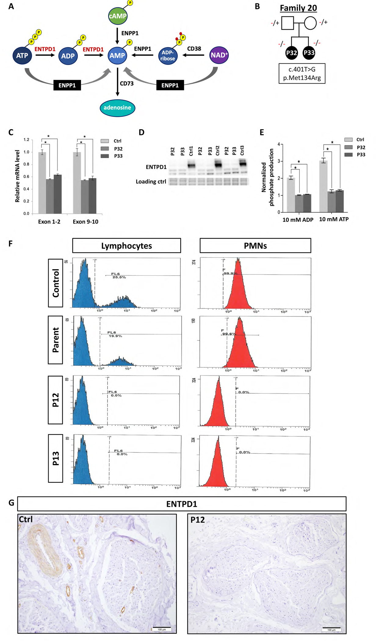
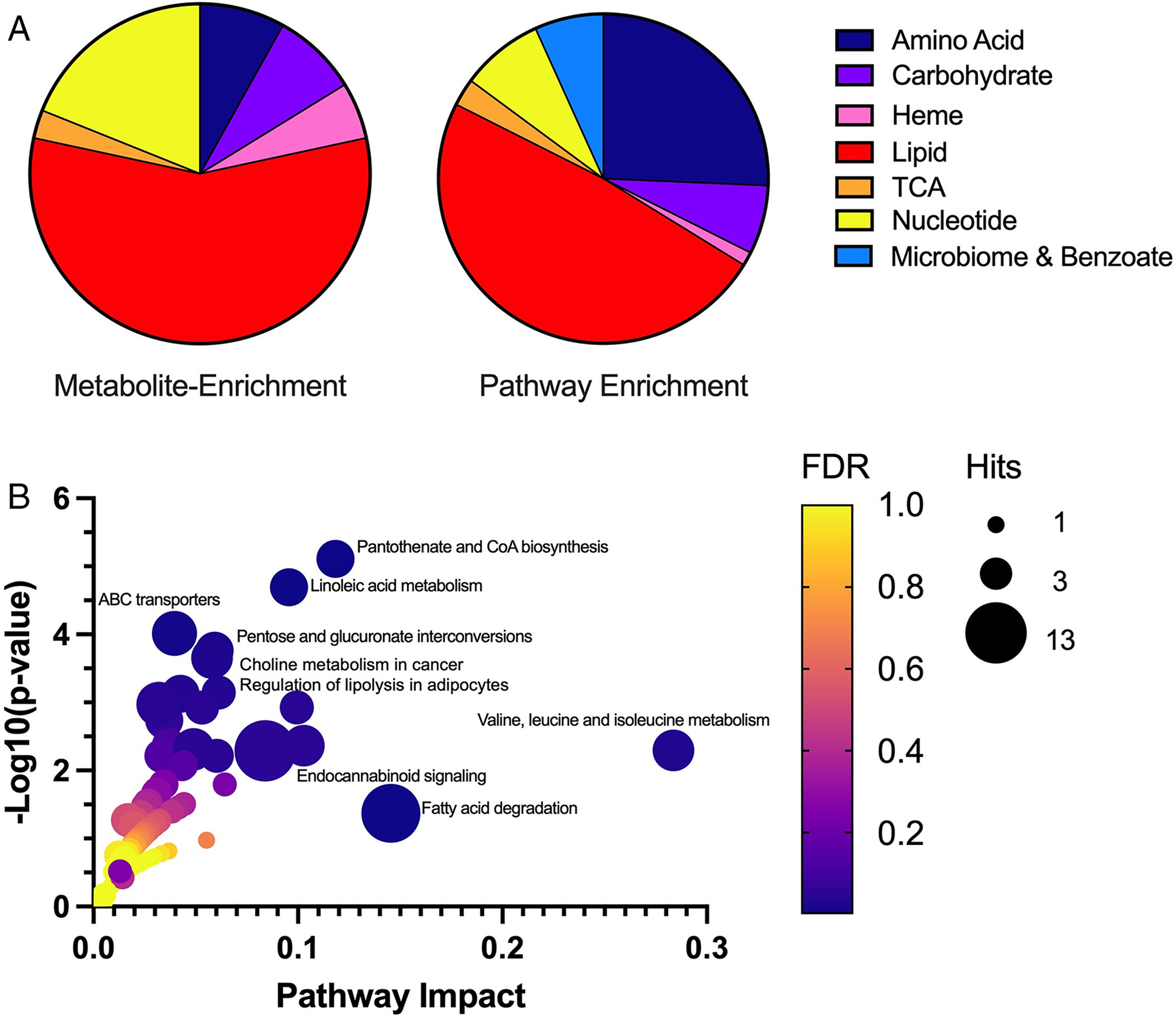
Results: A total of 27 individuals from 17 unrelated families were studied; additional phenotypic information was collected from published cases. Twelve novel pathogenic ENTPD1 variants are described (NM 001776.6): c.398_399delinsAA; p.(Gly133Glu), c.540del; p.(Thr181Leufs*18), c.640del; p.(Gly216Glufs*75), c.185 T > G; p.(Leu62*), c.1531 T > C; p.(*511Glnext*100), c.967C > T; p.(Gln323*), c.414-2_414-1del, and c.146 A > G; p.(Tyr49Cys) including 4 recurrent variants c.1109 T > A; p.(Leu370*), c.574-6_574-3del, c.770_771del; p.(Gly257Glufs*18), and c.1041del; p.(Ile348Phefs*19). Shared disease traits include childhood onset, progressive spastic paraplegia, intellectual disability (ID), dysarthria, and white matter abnormalities. In vitro assays demonstrate that ENTPD1 expression and function are impaired and that c.574-6_574-3del causes exon skipping. Global metabolomics demonstrate ENTPD1 deficiency leads to impaired nucleotide, lipid, and energy metabolism.

Interpretation: The ENTPD1 locus trait consists of childhood disease onset, ID, progressive spastic paraparesis, dysarthria, dysmorphisms, and white matter abnormalities, with some individuals showing neurocognitive regression. Investigation of an allelic series of ENTPD1 (1) expands previously described features of ENTPD1-related neurological disease, (2) highlights the importance of genotype-driven deep phenotyping, (3) documents the need for global collaborative efforts to characterize rare autosomal recessive disease traits, and (4) provides insights into disease trait neurobiology. ANN NEUROL 2022;92:304-321.


Conflict of interest statement

Conflicts of interests

J.R.L. has stock ownership in 23andMe, is a paid consultant for Regeneron Genetics Center, and is a co-inventor on multiple United States and European patents related to molecular diagnostics for inherited neuropathies, eye diseases, and bacterial genomic fingerprinting. The Department of Molecular and Human Genetics at Baylor College of Medicine receives revenue from clinical genetic testing conducted at Baylor Genetics (BG) Laboratories. M.C.K. is a paid consultant for PTC Therapeutics and Aeglea. Other authors have no potential conflicts to disclose.

Figures

Figure 1
Figure 1. Pedigrees and variant information for families with ENTPD1-related neurological disease.
(A) Pedigree and Sanger sequencing results of index family 1 with the homozygous variant NM_001766.6: c.398_399delinsAA; p.(Gly133Glu). (B) Absence of heterozygosity (AOH) plot of P1 showing a total AOH size of 310 Mb and 11.1 Mb of AOH around ENTPD1: c.398_399delinsAA (red line). (C) Conservation of amino acid residue p.Gly145 (D) Pedigrees and variant information of newly identified families with biallelic ENTPD1 variants and countries of origin.
Figure 2
Figure 2. Representative photographs and radiographs of individuals with ENTPD1-related neurological disease.
(A) Facial pictures of P17 at 7 years of age and P22 at 8 years showing low anterior hairline, synophrys, low-set ears with fleshy lobes, prominent philtrum, and micrognathia. (B) Pictures of P1 at 8 years of age and P22 at 8 years showing centripetal obesity, thoracic kyphosis, decreased lumbar lordosis, genu valgus, and cubitus valgus. (C) Representative hand images of P1, P6, and P10 at 8, 15, and 3 years, respectively, showing broad fingers with camptodactyly and spatulated finger tips (P1), mild camptodactyly of 4th and 5th digits (P6), and camptodactyly of all digits (P10). (D) Representative foot images of P1, P6, and P10 at 8, 15, and 3 years, respectively, showing broad toes with camptodactyly (P1), pes cavus with camptodactyly (P6), and broad great toes bilaterally and broad right 4th toe with camptodactyly (P10). (E) Hand radiographs of P1 at 8 years of age showing severe camptodactyly. (F) Foot radiographs of P1 at 8 years showing camptodactyly. (G) Foot radiographs of P6 at 15 years showing cavus and camptodactyly. (H) Foot radiographs of P28 at 19 years of age showing severe camptodactyly. (I) Sagittal spine radiograph of P1 at 8 years showing lumbar lordosis. (J) Hip radiograph of P10 at 3 years showing no gross abnormalities.
Figure 3
Figure 3. Individuals with biallelic pathogenic ENTPD1 variants have hypomyelination of the brain.
Representative magnetic resonance imaging (MRI) of the brain of affected individuals from different families at different ages. Arrows in the axial images are highlighting hypomyelination of the posterior limb of the internal capsule. (A) Sagittal T1-weighted imaging (1) and axial T2-FLAIR of P8 at 17 years of age. (B) and (C) Sagittal T1-weighted imaging (1) and axial T2-FLAIR of P9 at 3 and 4 years, respectively. (D) Sagittal T1-weighted imaging (1) and axial T2-FLAIR of P10 at 3 years. (E) Sagittal T2-weighted imaging (1) and axial T2-FLAIR of P17 at 7 years. (F) Sagittal T1-weighted imaging (1) and axial T2 of P29 at 15 years.
Figure 4
Figure 4. Biallelic pathogenic ENTPD1 variants identified in this cohort.
(A) Schematic showing chromosomal location and gene structure of ENTPD1. Previously unreported variants are labeled in red and previously published variants in black. (B) Linear amino acid structure of ENTPD1 and location of previously unreported (red) and published variant alleles (black).
Figure 5
Figure 5. Alternative splicing due to ENTPD1:c.574-6_574-3del results in skipping of exon 6.
(A) Schematic of ENTPD1 NM_001776.6, the most widely expressed trancript, showing 10 different exons. ENTPD1: c.574-6_574-3del is located in intron 5 (red asterisk). Arrows show location of primers for cDNA amplification. (B) Agarose gel electrophoresis image of ENTPD1 cDNA exon 4F and 7R amplification and schematic of resultant splicing products. Unaffected wildtype control cDNA and heterozygous parental cDNA show amplification of a bands at 818 bp not found in the affected proband sample (P7). Sanger sequencing confirmed that the 818 bp band contains exons 4, 5, 6, and 7 in control and unaffected parent. By contrast, the proband P7 who carries the homozygous ENTPD1: c.574-6_574-3del variant contains an alternative 572 bp product including exons 4, 5, and 7 only and thus skipping exon 6 completely. All three samples additionally contain a smaller product at 412 bp only containing exons 4 and 7. (C) Agarose gel electrophoresis image of ENTPD1 cDNA exon 6F and 10R amplification. Unaffected wildtype control cDNA and heterozygous parental cDNA show amplification of an 861 bp band containing exons 6, 7, 8, 9, and 10. No amplification is present in the homozygous proband sample.
Figure 6
Figure 6. Biallelic ENTPD1 variants impair ATP hydrolysis and ENTPD1 expression.
(A) ATP metabolic pathway showing the role of ENTPD1 in hydrolysis of ATP to ADP and ADP to AMP. (B) Lymphoblasts were derived from family 16 with two affected siblings (P27 and P28) with homozygous ENTPD1:c.401T>G; p.(Met134Arg) variant. (C) Reverse transcription-quantitative PCR of ENTPD1 mRNA levels, using primers spanning both exons 1–2 and exons 9–10 showing significantly decreased ENTPD1 mRNA levels in lymphoblasts from individuals with homozygous ENTPD1:c.401T>G variant. (D) Western blot of ENTPD1 p.(Met134Arg) showing deceased protein levels in patient lymphoblasts. The stain-free gel serves as the loading control. (E) Measured ATPase and ADPase activity using normalized phosphate production after incubation with either ATP or ADP at a final concentration of 10 mM for 30 min. * p<0.05. (F) Flow cytometry of ENTPD1+ lymphocytes and polymorphonuclear leukocytes (PMNs) in blood samples from P13 and P14 with homozygous ENTPD1:c.185T>G; p.(Leu62*) variant compared to control and heterozygous parental samples. (G) Immunohistochemical staining for ENTPD1 performed on paraffin sections of sural nerve biopsy from P13 with variant ENTPD1:c.185T>G shows complete absence of endo- and epineural vascular staining compared to control sample.
Figure 7
Figure 7
ENTPD1 deficiency alters multiple metabolic pathways important for lipid and energy metabolism. (A) Metabolite enrichment (left) illustrates molecule distribution of specific perturbed metabolites altered in at least two thirds of tested individuals (n = 37). Pathway enrichment (right) illustrates altered molecules from the same metabolic pathways (n = 148). In pathway enrichment, molecules may or may not be identical between patient samples; however, molecules fall within the same metabolic pathway. The distribution of these molecules across all patient samples illustrates the broader impact of lipid and energy metabolism due to ENTPD1 deficiency. (B) Gene–metabolite disease pathway interaction is shown for significantly perturbed metabolites in plasma of patients with ENTPD1 deficiency. Metabolites assessed (n = 98) are limited to molecules mapped to Kyoto Encyclopedia of Genes and Genomes pathways. The most significantly altered pathways (p < 0.05) are labeled. ABC = ATP-binding cassette; CoA = coenzyme A; TCA = tricarboxylic acid; FDR = false discovery rate.

References

    1. Lupski JR, Liu P, Stankiewicz P, Carvalho CMB, and Posey JE (2020). Clinical genomics and contextualizing genome variation in the diagnostic laboratory. Expert Rev Mol Diagn 20, 995–1002. - PMC - PubMed
    1. Karaca E, Posey JE, Coban Akdemir Z, Pehlivan D, Harel T, Jhangiani SN, Bayram Y, Song X, Bahrambeigi V, Yuregir OO, et al. (2018). Phenotypic expansion illuminates multilocus pathogenic variation. Genet Med 20, 1528–1537. - PMC - PubMed
    1. Tadahiro Mitani SI, Gezdirici Alper, Gulec Elif Yilmaz, Punetha Jaya, Fatih Jawid M., Herman Isabella, Gulsen Akay Tayfun Haowei Du, Calame Daniel G., Ayaz Akif, Tos Tulay, Yesil Gozde, Aydin Hatip, Geckinli Bilgen, Elcioglu Nursel, Candan Sukru, Sezer Ozlem, Haktan Bagis Erdem Davut Gul, Yasar Emine, Koparir Erkan, Elmas Muhsin, Yesilbas Osman, Kilic Betul, Serdal Güngör Ahmet C. Ceylan, Bozdogan Sevcan, Ture Mehmet, Etlik Ozdal, Cicek Salih, Aslan Huseyin, Yalcintepe Sinem, Topcu Vehap, Ipek Zeynep, Bayram Yavuz, Grochowski Christopher M., Jolly Angad, Dawood Moez, Duan Ruizhi, Jhangiani Shalini N., Doddapaneni Harsha, Hu Jianhong, Muzny Donna M., Baylor-Hopkins Center for Mendelian Genomics, Marafi Dana, Akdemir Zeynep Coban, Karaca Ender, Carvalho Claudia MBC, Gibbs Richard A., Posey Jennifer E., Lupski James R., and Pehlivan Davut. (2021). Evidence for a higher prevalence of oligogenic inheritance in neurodevelopmental disorders in the Turkish population. American Journal of Human Genetics. - PMC - PubMed
    1. Ruano L, Melo C, Silva MC, and Coutinho P (2014). The global epidemiology of hereditary ataxia and spastic paraplegia: a systematic review of prevalence studies. Neuroepidemiology 42, 174–183. - PubMed
    1. Blackstone C (2018). Hereditary spastic paraplegia. Handb Clin Neurol 148, 633–652. - PubMed