. 2022 Apr 26;18(4):e1009962.
doi: 10.1371/journal.pcbi.1009962. eCollection 2022 Apr.

Multi-targeting of K-Ras domains and mutations by peptide and small molecule inhibitors

Affiliations

Multi-targeting of K-Ras domains and mutations by peptide and small molecule inhibitors

Mansour Poorebrahim et al. PLoS Comput Biol. .

Abstract

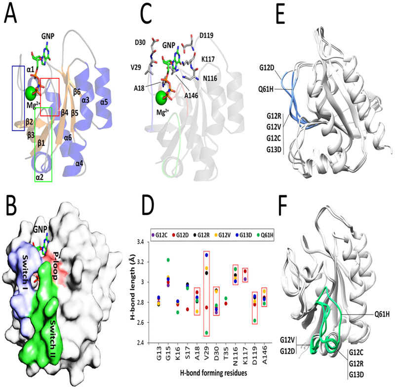
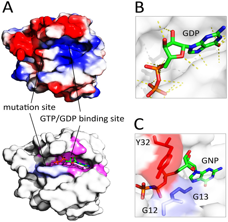
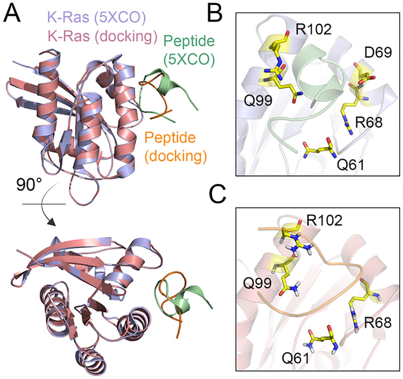
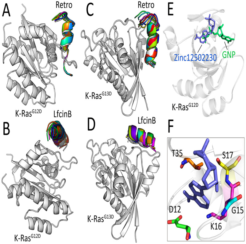
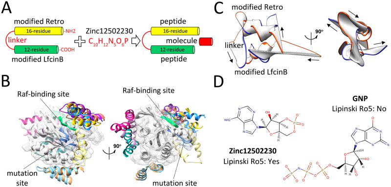
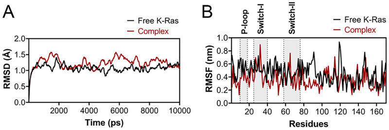
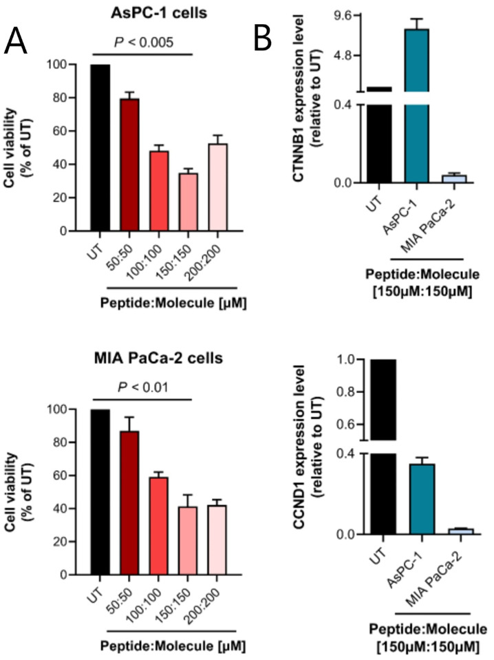
K-Ras activating mutations are significantly associated with tumor progression and aggressive metastatic behavior in various human cancers including pancreatic cancer. So far, despite a large number of concerted efforts, targeting of mutant-type K-Ras has not been successful. In this regard, we aimed to target this oncogene by a combinational approach consisting of small peptide and small molecule inhibitors. Based on a comprehensive analysis of structural and physicochemical properties of predominantly K-Ras mutants, an anti-cancer peptide library and a small molecule library were screened to simultaneously target oncogenic mutations and functional domains of mutant-type K-Ras located in the P-loop, switch I, and switch II regions. The selected peptide and small molecule showed notable binding affinities to their corresponding binding sites, and hindered the growth of tumor cells carrying K-RasG12D and K-RasG12C mutations. Of note, the expression of K-Ras downstream genes (i.e., CTNNB1, CCND1) was diminished in the treated Kras-positive cells. In conclusion, our combinational platform signifies a new potential for blockade of oncogenic K-Ras and thereby prevention of tumor progression and metastasis. However, further validations are still required regarding the in vitro and in vivo efficacy and safety of this approach.


Conflict of interest statement

The authors have declared that no competing interests exist.

Figures

Fig 1
Fig 1. Assessment of nucleotide-binding site of K-Ras bound to GTP/GDP.
(A) Cartoon representation of K-Ras structure bound to GNP (PDB ID: 2PMX). The β-strands, α-helices, and coils are shown in orange, blue, and gray colors, respectively. The P-loop, switch I, and switch II domains are indicated in red, blue, and green boxes, respectively. (B) Surface representation of GTP-bound K-Ras along with its nucleotide-binding sites. (C) Arrangements of the 7-residue binding interface in the GTP-bound K-Ras. (D) Calculation of H-bond distances between the atoms of 12 common nucleotide-binding residues and GTP/GDP in all studied K-Ras mutants. The H-bond length of 7 residues located outside of the previously characterized GTP/GDP-binding site is shown in red boxes. (E) Effects of the oncogenic mutations on switch I (blue) and (F) switch II (green) domains of K-Ras mutants.
Fig 2
Fig 2. Evaluation of surface charge pattern of the K-RasG12D.
(A) Calculation of the electrostatic surface potential of wild-type K-Ras. The mutation sites 12, 13, and 61 (blue in the below figure) and GTP/GDP binding sites (magenta in the below figure) of the protein are shown with solid lines. The blue, red, and gray colors in the above figure refer to the positively-charged, negatively-charged, and hydrophobic regions, respectively (B) Polar contacts (yellow dotted lines) between GDP and K-RasG12D. (C) A close-up view of the closed conformation of the wild-type GTP-bound K-Ras through direct interaction of G12/13 with Y32.
Fig 3
Fig 3. Validation of molecular docking method.
(A) Structural alignment of the K-RasG12D-peptide complex determined by X-ray crystallography (ID: 5XCO) and the docking model. Close-up view of the binding interface between K-RasG12D and inhibitory peptide in the (B) crystallography and (C) docking studies. The residues of K-RasG12D forming H-bond interactions with the peptide are shown in yellow.
Fig 4
Fig 4. Interaction mode of peptides/molecule with K-RasG12D and K-RasG13D.
Superimposition of top-ranked peptide-protein docked poses resulted from flexible docking of (A) the modified Retro-KrasG12D, (B) the modified LfcinB- KrasG12D, (C) the modified Retro-KrasG13D, and (D) the modified LfcinB-KrasG13D. (E) Superimposition of GNP-bound (green) and Zinc12502230-bound (blue) K-RasG12D. (F) Close-up view of H-bonding interactions between Zinc12502230 (blue) and five residues of K-RasG12D colored in different colors.
Fig 5
Fig 5. Binding energy and structural evaluations of final selected peptides and molecule.
(A) Schematic representation of dimer peptide construction and combination of this peptide with Zinc12502230. (B) Ten top-ranked peptide-protein complexes obtained from flexible docking of the modified Retro-LfcinB peptide (each pose in a different color) with K-RasG12D (gray). The Raf-binding and mutation sites are shown in green and blue colors, respectively. (C) The dominant mode of motion resulted from 50 frames of Retro-LfcinB MD simulations. The orange and blue colors represent the first and last frames, respectively. Arrows show the direction of motions. (D) Molecular representation of the small molecule Zinc12502230 (left) and GNP (right).
Fig 6
Fig 6. RMSD and RMSF plots of free K-Ras and Kras-peptide complex.
(A) The RMSD and (B) RMSF of plots of free (black) and complex (red) K-Ras forms after a 10 ns MD simulation. The position of target domains is shown the B panel.
Fig 7
Fig 7. MTT assay and quantitative real-time PCR results.
(A) Assessment of the cell viability of AsPC-1 (up) and MIA PaCa-2 (down) cells after treatment with different concentrations of Retro-LfcinB peptide and Zinc12502230 small molecule. Values were normalized with untreated cells. The highest cytotoxicity of the compounds (at 150μM of peptide: 150μM of the small molecule) was significantly different from untreated control at P < 0.005 and P < 0.01 (Student’s t-test) in AsPC-1 and MIA PaCa-2 cells, respectively. UT: untreated cells. (B) Cancer cells expressing K-RasG12D were treated with the combination of 150μM of modified Retro-LfcinB peptide and 150μM of Zinc12502230 small molecule during 72 h, and then mRNA level was analyzed with quantitative real-time PCR for expression of K-Ras down-stream genes including CTNNB1 and CCND1. Results are the mean ± SE of three separate experiments.

References

    1. Pylayeva-Gupta Y, Grabocka E, Bar-Sagi D. RAS oncogenes: weaving a tumorigenic web. Nature reviews Cancer. 2011;11(11):761–74. Epub 2011/10/14. doi: 10.1038/nrc3106 . - DOI - PMC - PubMed
    1. Wang Y, Kaiser CE, Frett B, Li H-y. Targeting mutant KRAS for anticancer therapeutics: a review of novel small molecule modulators. Journal of medicinal chemistry. 2013;56(13):5219–30. doi: 10.1021/jm3017706 - DOI - PMC - PubMed
    1. Lemieux E, Cagnol S, Beaudry K, Carrier J, Rivard N. Oncogenic KRAS signalling promotes the Wnt/β-catenin pathway through LRP6 in colorectal cancer. Oncogene. 2015;34(38):4914–27. Epub 2014/12/17. doi: 10.1038/onc.2014.416 . - DOI - PMC - PubMed
    1. Downward J. Targeting RAS signalling pathways in cancer therapy. Nature reviews Cancer. 2003;3(1):11–22. Epub 2003/01/02. doi: 10.1038/nrc969 . - DOI - PubMed
    1. Adjei AA. Blocking oncogenic Ras signaling for cancer therapy. Journal of the National Cancer Institute. 2001;93(14):1062–74. Epub 2001/07/19. doi: 10.1093/jnci/93.14.1062 . - DOI - PubMed

MeSH terms