. 2022 Apr 25;57(8):959-973.e7.
doi: 10.1016/j.devcel.2022.03.017.

RBPMS is an RNA-binding protein that mediates cardiomyocyte binucleation and cardiovascular development

Affiliations

RBPMS is an RNA-binding protein that mediates cardiomyocyte binucleation and cardiovascular development

Peiheng Gan et al. Dev Cell. .

Abstract

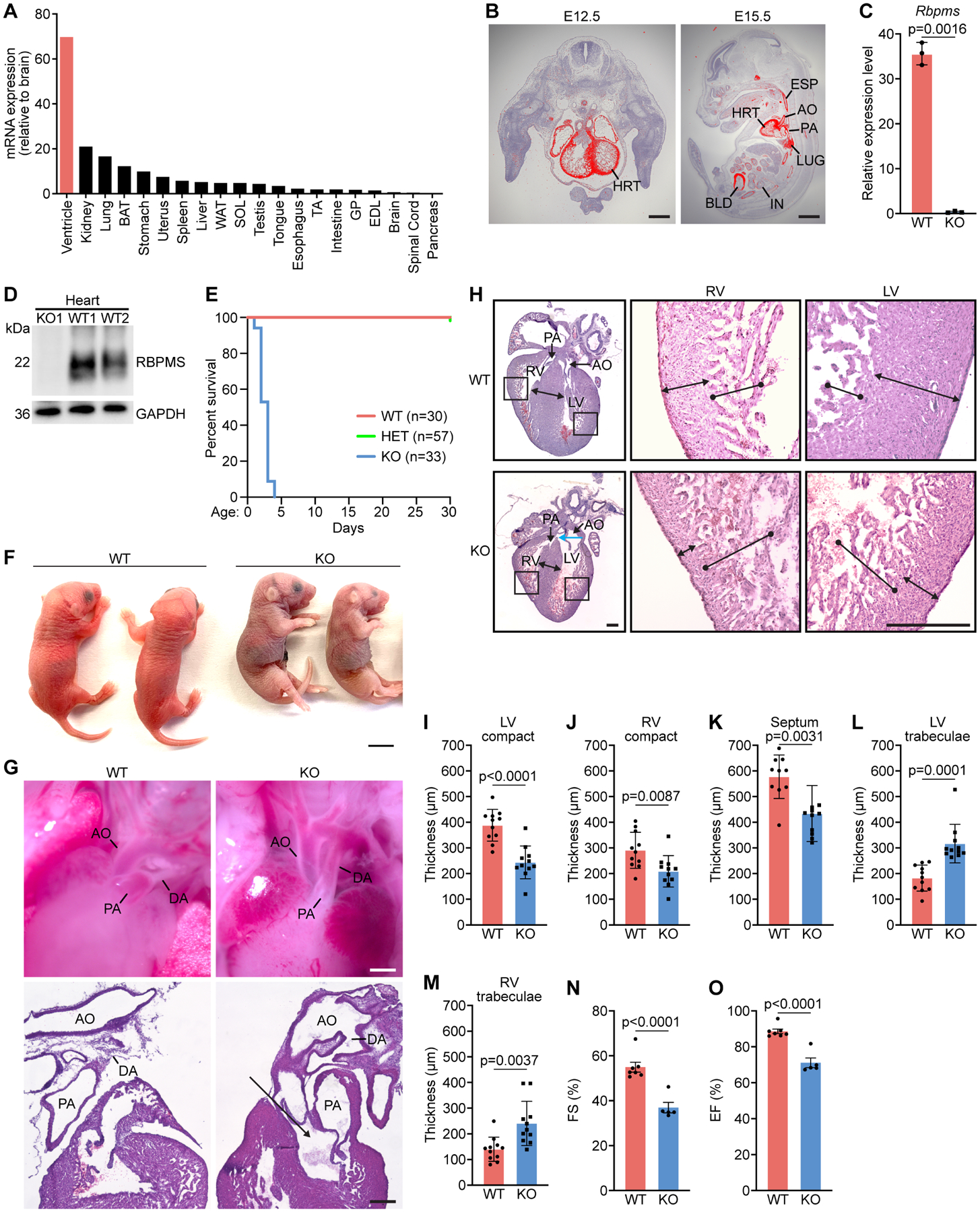
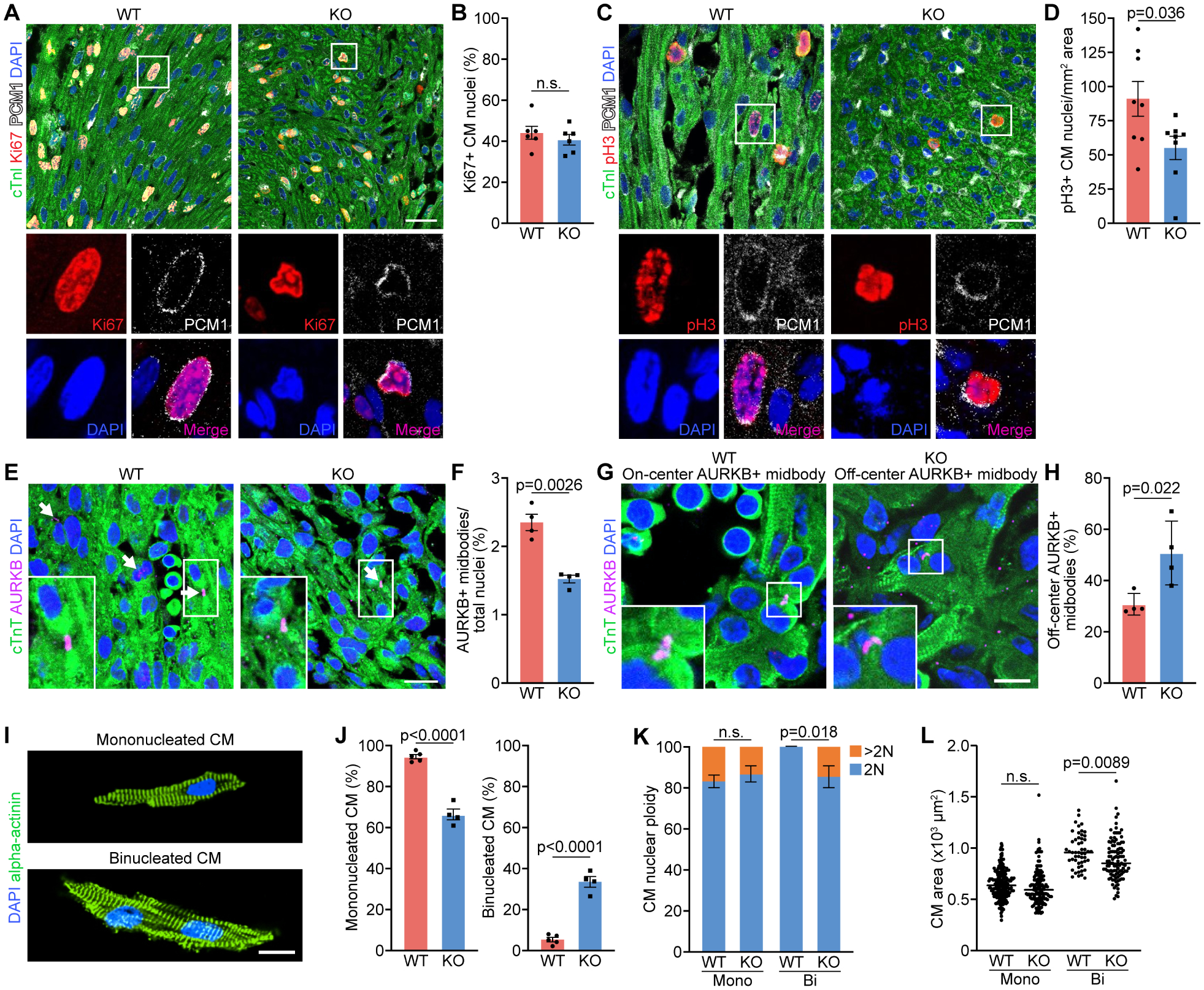
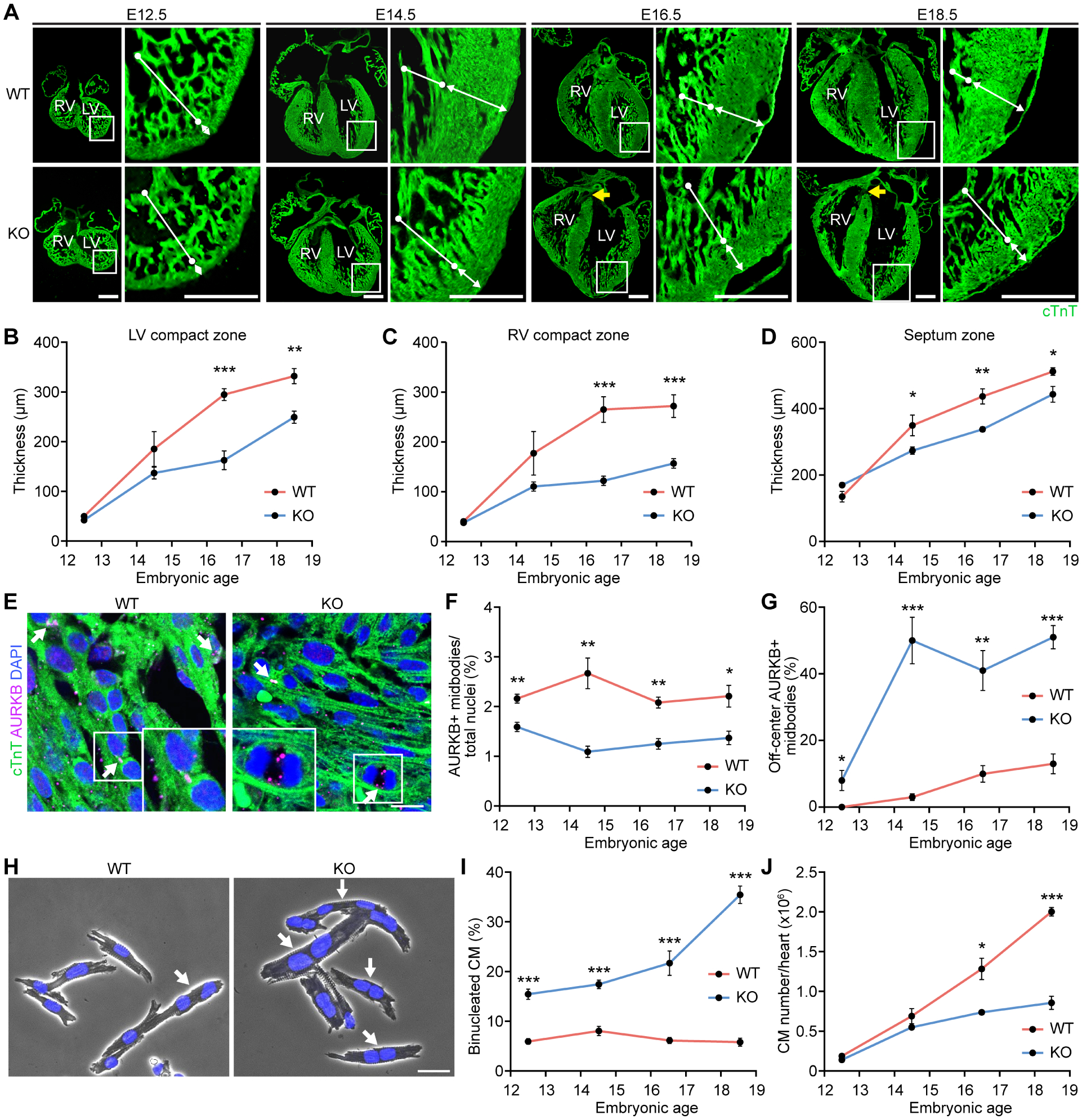
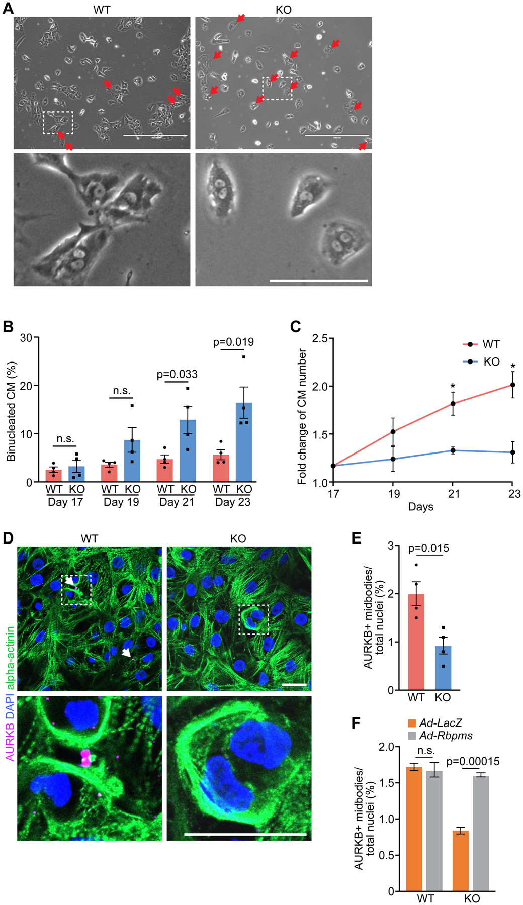
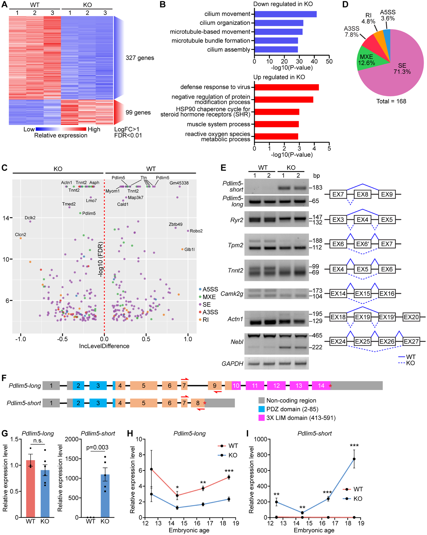
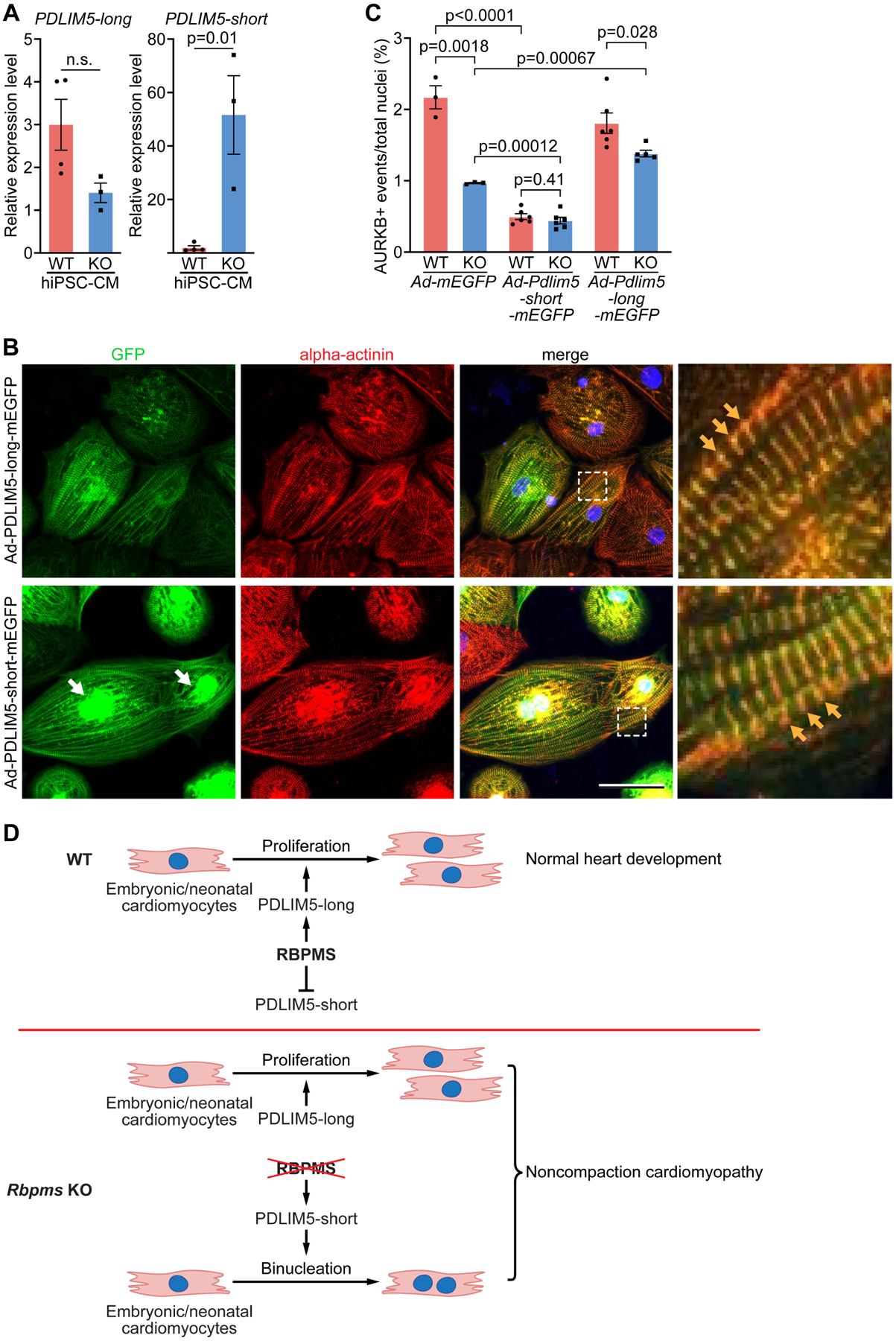
Noncompaction cardiomyopathy is a common congenital cardiac disorder associated with abnormal ventricular cardiomyocyte trabeculation and impaired pump function. The genetic basis and underlying mechanisms of this disorder remain elusive. We show that the genetic deletion of RNA-binding protein with multiple splicing (Rbpms), an uncharacterized RNA-binding factor, causes perinatal lethality in mice due to congenital cardiovascular defects. The loss of Rbpms causes premature onset of cardiomyocyte binucleation and cell cycle arrest during development. Human iPSC-derived cardiomyocytes with RBPMS gene deletion have a similar blockade to cytokinesis. Sequencing analysis revealed that RBPMS plays a role in RNA splicing and influences RNAs involved in cytoskeletal signaling pathways. We found that RBPMS mediates the isoform switching of the heart-enriched LIM domain protein Pdlim5. The loss of Rbpms leads to an abnormal accumulation of Pdlim5-short isoforms, disrupting cardiomyocyte cytokinesis. Our findings connect premature cardiomyocyte binucleation to noncompaction cardiomyopathy and highlight the role of RBPMS in this process.

Keywords: Pdlim5; RNA-binding protein; Rbpms; alternative splicing; cardiomyocyte binucleation; hypertrabeculation; noncompaction cardiomyopathy; patent ductus arteriosus.


Conflict of interest statement

Declaration of interests E.N.O. is on the editorial board of Developmental Cell. The authors declare no other competing interests.

Figures

Figure 1.
Figure 1.. Perinatal lethality and noncompaction cardiomyopathy of Rbpms KO mice.
A. Relative expression levels of Rbpms mRNA in adult mouse tissues as measured by qRT-PCR. B. In situ hybridization showing cardiac and smooth muscle expression of Rbpms mRNA at the indicated embryonic time points. HRT, heart; AO: aorta; PA: pulmonary artery; IN, intestine; LUG, lung; BLD, bladder; ESP, esophagus. Scale bars (left): 500 μm, (right): 2 mm. C. Expression level of Rbpms mRNA in P1 WT and KO hearts, measured by RNA-seq (n = 3 for WT, and 3 for KO). D. Western blot analysis showing loss of RBPMS protein in P1 hearts of Rbpms KO mice. GAPDH is a loading control. E. Survival curve of Rbpms KO mice. F. Representative image of Rbpms WT and KO pups at P2. Scale bar: 5 mm. G. Whole mount (top panel) and H&E sagittal sections showing the ductus arteriosus (DA), pulmonary artery (PA), aorta artery (AO) in WT and KO pups 6 h after birth. Black arrow indicates double Outlet Right Ventricle (DORV) in KO heart. Scale bars (top): 500 μm, (bottom): 200 μm. H. H&E-stained coronal sections of representative hearts from P1 WT and KO pups fixed in diastole. Magnified images show left and right ventricular regions. Blue arrow in KO heart section indicates overriding aorta. Scale bar: 300 μm. I-M. Measurements of left ventricle (LV) compact myocardium, right ventricle (RV) compact myocardium, septal, LV trabecular and RV trabecular zone thicknesses in coronal sections at the level of papillary muscle roots (n = 11 for WT and KO). N, O. Fractional shortening (FS%) and ejection fraction (EF%) of WT and KO mouse hearts between P1–P3 (n = 6 for WT, and 5 for KO). All data are presented as mean ± SEM.
Figure 2.
Figure 2.. Cytokinesis defects of P1 Rbpms KO mouse cardiomyocytes.
A, B. Immunofluorescence staining for Ki67, cardiac troponin I (cTnI), PCM1 and DAPI on coronal sections of WT and KO myocardium at P1 and quantification of percentage of Ki67+ cardiomyocyte (CM) nuclei over total nuclei (n = 6 for WT and KO). Scale bar: 30 μm. C, D. Immunofluorescence staining for phosphorylated histone H3 (pH3), cTnI, PCM1 and DAPI of WT and KO myocardium at P1, and quantification of number of pH3+ CM nuclei per mm2 area. (n = 8 for WT and KO). Scale bar: 30 μm. E, F. Immunofluorescence staining for aurora B kinase (AURKB), cTnT and DAPI of WT and KO hearts at P1 and quantification of AURKB-positive midbody frequency (n = 4 for WT and KO). Scale bar: 25 μm. White arrows indicate AURKB-positive midbodies between nuclei. G. Representative immunofluorescence staining images of on-center and off-center AURKB-positive midbodies in P1 WT and KO heart sections. Scale bar: 10 μm. H. Percentage of off-center AURKB-positive midbodies in WT and KO hearts at P1 (n = 4 for WT and KO). I. Representative immunofluorescence images of P1 mononucleated and binucleated CMs stained for alpha-actinin. Scale bar: 20 μm. J. Percentage of mononucleated and binucleated CMs of P1 WT and KO hearts (n = 5 for WT, and 4 for KO). K. Quantification of the nuclear ploidy of the mononucleated (mono) and binucleated (Bi) CMs in P1 WT and KO hearts (n = 5 for WT mice, and 4 for KO mice, average 100–200 cardiomyocytes per mouse). n.s., not significant. L. Areas of individual mononucleated and binucleated CMs (n = 5 for WT mice, and 4 for KO mice, average 100–200 cardiomyocytes per mouse). n.s., not significant. All data are presented as mean ± SEM.
Figure 3.
Figure 3.. Noncompaction cardiomyopathy in Rbpms KO mice is a developmental defect.
A. Representative immunofluorescence images of coronal heart sections at indicated embryonic time points stained for cTnT. White bars with round head indicate LV trabeculae, and white bars with arrowhead indicate LV compact zone. Yellow arrows in KO heart sections indicate overriding aorta. Scale bar: 400 μm. B-D. Quantification of thickness of LV compact zone, RV compact zone and septum zone in embryonic hearts (n = 5–7 for WT and KO at each time point). E. Representative immunofluorescence images of WT and KO hearts at E16. 5 stained for AURKB and cTnT. Scale bar: 25 μm. White arrows indicate AURKB-positive midbodies between nuclei. F. Quantification of AURKB-positive midbody frequency in embryonic hearts (n = 5–7 for WT and KO at each time point). G. Quantification of off-center AURKB-positive midbody percentage in embryonic hearts (n = 4–6 for WT and KO at each time point). H. Representative phase contrast microscopy images of isolated cardiomyocytes from E18.5 WT and KO hearts stained with DAPI. Scale bar: 30 μm. White arrows indicate binucleated cardiomyocytes. I. Quantification of binucleated cardiomyocytes in embryonic hearts (n = 3–6 for WT and KO mice at each time point, average 100–200 cardiomyocytes per mouse). J. Total cardiomyocyte number in hearts at the indicated embryonic time points (n = 5–7 for WT and KO at each time point). *p<0.05, **p<0.01; ***p<0.001; n.s., not significant. All data are presented as mean ± SEM.
Figure 4.
Figure 4.. Cytokinesis defects in RBPMS-KO hiPSC-derived cardiomyocytes.
A. Phase contrast microscopy of WT and RBPMS-KO hiPSC-cardiomyocytes. Red arrows indicate binucleated hiPSC-cardiomyocytes. Scale bars (top): 500 μm, (bottom): 100 μm. B. Percentage of binucleated WT and RBPMS-KO hiPSC-cardiomyocytes at indicated time points (n = 3 for WT groups, and 4 for KO groups, average 200 cardiomyocytes per group). n.s., not significant. C. Fold change of cardiomyocytes number in WT and RBPMS-KO cardiomyocytes at indicated time points. D. Immunofluorescent staining for AURKB and alpha-actinin in WT and RBPMS-KO hiPSC-cardiomyocytes. White arrows indicate AURKB+ midbodies in between nuclei. Scale bar: 25 μm. E. Quantification of AURKB-positive midbody frequencies in WT and RBPMS-KO hiPSC-cardiomyocytes (n = 4 for WT and KO groups, average 300 cardiomyocytes per group). F. Quantification of AURKB-positive midbody frequencies in WT and RBPMS-KO hiPSC-cardiomyocytes infected with Ad-LacZ and Ad-Rbpms (n = 4 for WT groups, and 3 for KO groups, average 300 cardiomyocytes per group). All data are presented as mean ± SEM.
Figure 5.
Figure 5.. Paired-end RNA sequencing reveals Pdlim5 as a splicing target of RBPMS.
A. Heatmap of differentially expressed genes between WT and KO P1 heart ventricles identified by RNA-seq. B. Top GO terms for down and upregulated genes in KO samples. C. Volcano plot showing the inclusion (Inc) level differences (WT Inclevel – KO Inclevel, X axis) and False Discovery Rates (FDR) for differential alternative splicing events (ASEs) between WT and KO P1 heart ventricles. Y axis is presented as −log10(FDR). D. Pie chart showing the percentage of different ASEs in Rbpms KO mice (FDR<0.01). SE: skipped exon, MXE: mutually exclusive exon, RI: retained intron, A3SS: alternative 3’ splice site, and A5SS: alternative 5’ splice site. E. Representative RT-PCR confirmation of abnormal splicing events in KO heart, and schematic diagram of SE events based on rMATs analysis. Solid lines indicate normal splicing events, and dashed lines indicate abnormal events in KO hearts. F. Schematic of murine Pdlim5 long and short isoform gene structures. Boxes represent exons, different functional domains are labelled with different colors. Red arrows indicate the locations of qRT-PCR primers for determining long and short isoforms expression. G. qRT-PCR of the relative expression of Pdlim5 long and short isoforms in P1 WT and KO heart (n = 3 for WT, and 6 for KO). H, I. qRT-PCR of relative expression levels of Pdlim5-long and short isoforms in WT and KO hearts at different embryonic time points (n = 4–6 for WT or KO). *p<0.05, **p<0.01; ***p<0.001; n.s., not significant. All data are presented as mean ± SEM.
Figure 6.
Figure 6.. Pdlim5 long and short variants regulate cytokinesis in hiPSC-cardiomyocytes.
A. Quantification of relative expression levels of PDLIM5 long and short isoforms in WT and KO hiPSC-cardiomyocytes by qRT-PCR (n = 4 for WT groups, and 3 for KO groups). B. hiPSC-cardiomyocytes were infected with Ad-Pdlim5-long-mEGFP and Ad-Pdlim5-short-mEGFP and immunostained for GFP and alpha-actinin. White arrows indicate the accumulation of Pdlim5-short variants surrounding nuclei. Yellow arrows indicate Pdlim5 long and short isoforms colocalizing with alpha-actinin at Z-discs. Scale bar: 50 μm C. Quantification of AURKB-positive midbody frequency in WT and KO hiPSC-cardiomyocytes infected with Ad-mEGFP, Ad-Pdlim5-long-mEGFP and Ad-Pdlim5-short-mEGFP (n = 4–6 for WT or KO groups, average 300 cardiomyocytes per group). D. Schematic diagrams showing the function of RBPMS in cardiomyocyte proliferation and WT heart development (top), and loss of Rbpms causes cardiomyocyte binucleation and noncompaction cardiomyopathy (bottom). All data are presented as mean ± SEM.

Comment in

References

    1. AKERBERG AA, BURNS CE & BURNS CG 2019. Exploring the Activities of RBPMS Proteins in Myocardial Biology. Pediatr Cardiol, 40, 1410–1418. - PMC - PubMed
    1. ALI H, BRAGA L & GIACCA M 2020. Cardiac regeneration and remodelling of the cardiomyocyte cytoarchitecture. FEBS J, 287, 417–438. - PubMed
    1. ALMEIDA AG & PINTO FJ 2013. Non-compaction cardiomyopathy. Heart, 99, 1535–42. - PubMed
    1. ARNDT AK, SCHAFER S, DRENCKHAHN JD, SABEH MK, PLOVIE ER, CALIEBE A, KLOPOCKI E, MUSSO G, WERDICH AA, KALWA H, HEINIG M, PADERA RF, WASSILEW K, BLUHM J, HARNACK C, MARTITZ J, BARTON PJ, GREUTMANN M, BERGER F, HUBNER N, SIEBERT R, KRAMER HH, COOK SA, MACRAE CA & KLAASSEN S 2013. Fine mapping of the 1p36 deletion syndrome identifies mutation of PRDM16 as a cause of cardiomyopathy. Am J Hum Genet, 93, 67–77. - PMC - PubMed
    1. BERGMANN O, BHARDWAJ RD, BERNARD S, ZDUNEK S, BARNABE-HEIDER F, WALSH S, ZUPICICH J, ALKASS K, BUCHHOLZ BA, DRUID H, JOVINGE S & FRISEN J 2009. Evidence for cardiomyocyte renewal in humans. Science, 324, 98–102. - PMC - PubMed

Publication types

LinkOut - more resources