

Recent advances and clinical applications of deep learning in medical image analysis
- PMID: 35472844
- PMCID: PMC9156578
- DOI: 10.1016/j.media.2022.102444
Recent advances and clinical applications of deep learning in medical image analysis
Abstract
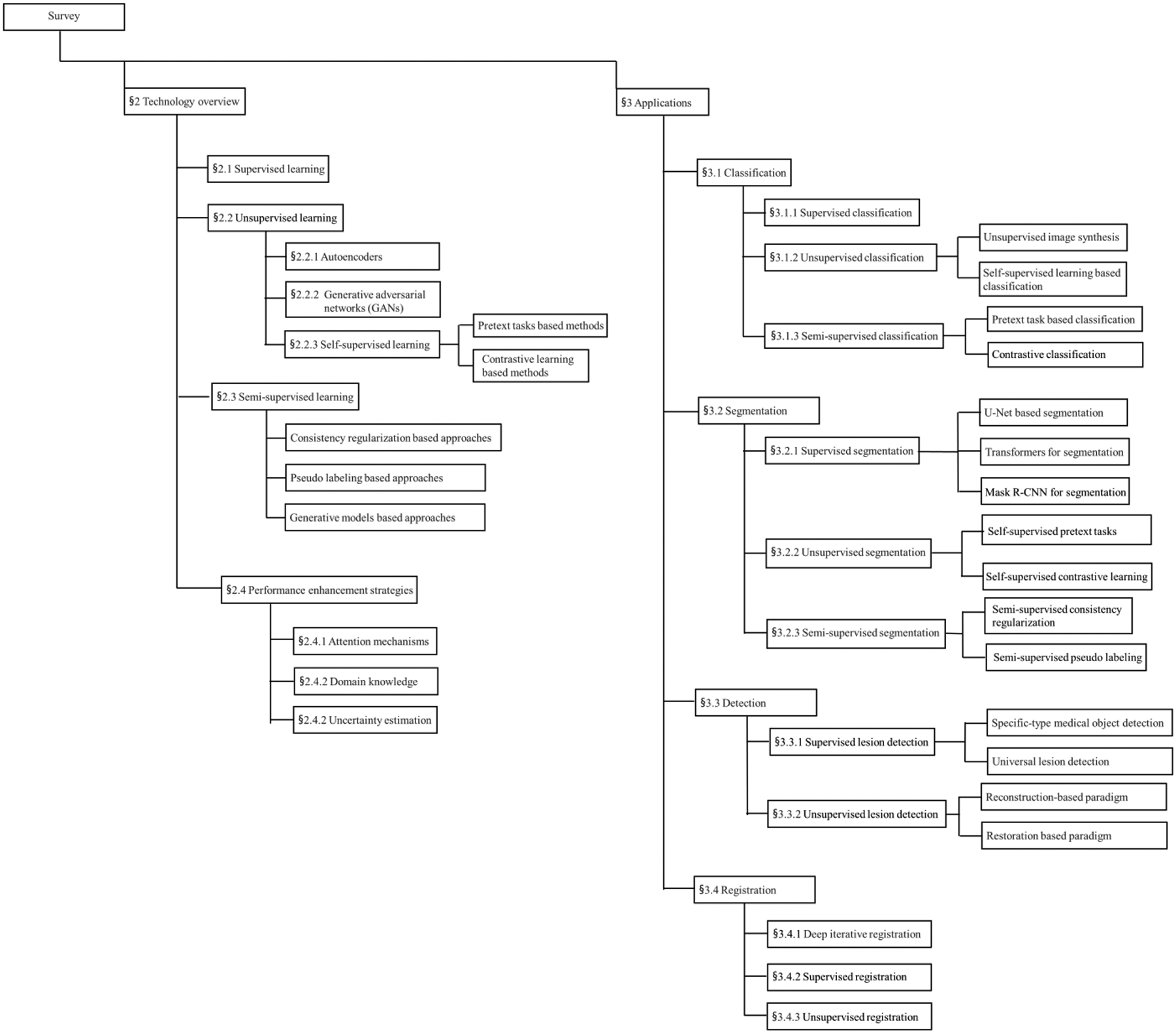
Deep learning has received extensive research interest in developing new medical image processing algorithms, and deep learning based models have been remarkably successful in a variety of medical imaging tasks to support disease detection and diagnosis. Despite the success, the further improvement of deep learning models in medical image analysis is majorly bottlenecked by the lack of large-sized and well-annotated datasets. In the past five years, many studies have focused on addressing this challenge. In this paper, we reviewed and summarized these recent studies to provide a comprehensive overview of applying deep learning methods in various medical image analysis tasks. Especially, we emphasize the latest progress and contributions of state-of-the-art unsupervised and semi-supervised deep learning in medical image analysis, which are summarized based on different application scenarios, including classification, segmentation, detection, and image registration. We also discuss major technical challenges and suggest possible solutions in the future research efforts.
Keywords: Attention; Classification; Deep learning; Detection; Medical images; Registration; Segmentation; Self-supervised learning; Semi-supervised learning; Unsupervised learning; Vision Transformer.
Copyright © 2022. Published by Elsevier B.V.
Conflict of interest statement
Declaration of Competing Interest The authors declare that they have no known competing financial interests or personal relationships that could have appeared to influence the work reported in this paper.
Figures

References
-
- Meyers PH, Nice CM Jr, Becker HC, Nettleton WJ Jr, Sweeney JW, Meckstroth GR, 1964. Automated computer analysis of radiographic images. Radiology 83, 1029–1034. - PubMed
-
- Kruger RP, Townes JR, Hall DL, Dwyer SJ, Lodwick GS, 1972. Automated Radiographic Diagnosis via Feature Extraction and Classification of Cardiac Size and Shape Descriptors. IEEE Transactions on Biomedical Engineering BME-19, 174–186. - PubMed
-
- Sezaki N, Ukena K, 1973. Automatic Computation of the Cardiothoracic Ratio with Application to Mass Screening. IEEE Transactions on Biomedical Engineering BME-20, 248–253. - PubMed
-
- Doi K, MacMahon H, Katsuragawa S, Nishikawa RM, Jiang Y, 1999. Computer-aided diagnosis in radiology: potential and pitfalls. European Journal of Radiology 31, 97–109. - PubMed
Publication types
MeSH terms
Grants and funding
LinkOut - more resources
Full Text Sources
Medical
Miscellaneous
