. 2022 Apr 26;14(1):59.
doi: 10.1186/s13195-022-00999-5.

Loss of perivascular aquaporin-4 localization impairs glymphatic exchange and promotes amyloid β plaque formation in mice

Affiliations

Loss of perivascular aquaporin-4 localization impairs glymphatic exchange and promotes amyloid β plaque formation in mice

Matthew Simon et al. Alzheimers Res Ther. .

Abstract

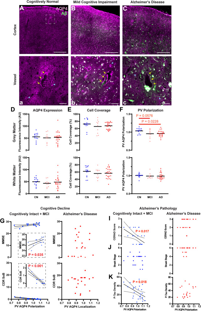
Background: Slowed clearance of amyloid β (Aβ) is believed to underlie the development of Aβ plaques that characterize Alzheimer's disease (AD). Aβ is cleared in part by the glymphatic system, a brain-wide network of perivascular pathways that supports the exchange of cerebrospinal and brain interstitial fluid. Glymphatic clearance, or perivascular CSF-interstitial fluid exchange, is dependent on the astroglial water channel aquaporin-4 (AQP4) as deletion of Aqp4 in mice slows perivascular exchange, impairs Aβ clearance, and promotes Aβ plaque formation.

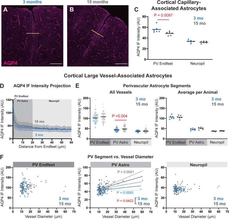
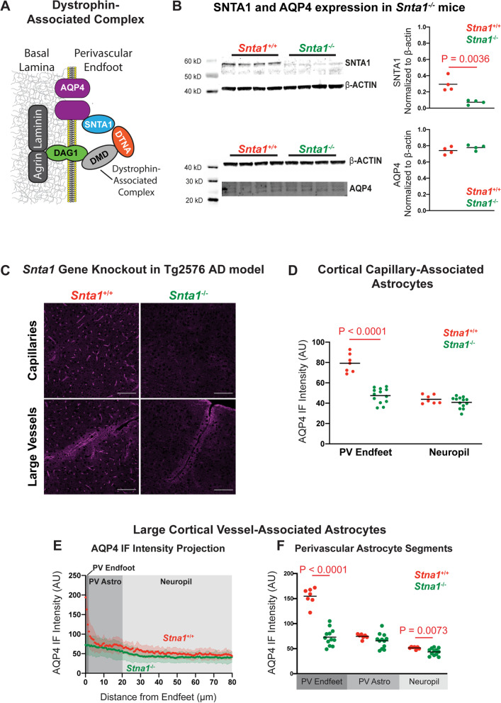
Methods: To define the role of AQP4 in human AD, we evaluated AQP4 expression and localization in a human post mortem case series. We then used the α-syntrophin (Snta1) knockout mouse model which lacks perivascular AQP4 localization to evaluate the effect that loss of perivascular AQP4 localization has on glymphatic CSF tracer distribution. Lastly, we crossed this line into a mouse model of amyloidosis (Tg2576 mice) to evaluate the effect of AQP4 localization on amyloid β levels.

Results: In the post mortem case series, we observed that the perivascular localization of AQP4 is reduced in frontal cortical gray matter of subjects with AD compared to cognitively intact subjects. This decline in perivascular AQP4 localization was associated with increasing Aβ and neurofibrillary pathological burden, and with cognitive decline prior to dementia onset. In rodent studies, Snta1 gene deletion slowed CSF tracer influx and interstitial tracer efflux from the mouse brain and increased amyloid β levels.

Conclusions: These findings suggest that the loss of perivascular AQP4 localization may contribute to the development of AD pathology in human populations.

Keywords: AQP4; Alzheimer’s disease; Amyloid β; Aquaporin-4; Astrocyte; Perivascular; glymphatic; α-Syntrophin.


Conflict of interest statement

The neuropathological case series included in this study was funded through a Sponsored Collaborative Agreement with GlaxoSmithKline to JJI. The other authors declare that they have no competing interests.

Figures

Fig. 1
Fig. 1
Loss of perivascular AQP4 localization is associated with pathology and cognitive impairment in the early stage Alzheimer’s disease. A–C, a–c Representative images of the immunofluorescent labeling of AQP4 and Aβ in frontal cortex of cognitively normal (CN, A, a), mild cognitive impairment (MCI, B, b), and Alzheimer’s disease (AD, C, c) subjects. The bottom panel (a–c) shows the areas indicated by white squares in the top panel (A–C) at higher magnification. Yellow arrows indicated the large vessels and blue arrow heads indicate the microvessels. Scale bar: 1 mm (A–C) and 0.1 mm (a–c). In CN subjects, AQP4 expression was largely uniform through the cortical layers between the pial surface and subcortical white matter boundary (A), with increased IF at perivascular endfeet surrounding the cerebral vasculature (a). In MCI and AD subjects, AQP4 expression became more irregular (B–C, b–c). Quantification of mean AQP4 IF (D) and cell coverage (E) showed comparable values between CN, MCI, and AD groups frontal cortical grey matter (top) and white matter (bottom). F Perivascular polarization was significantly reduced in AD compared to CN subjects in frontal cortical gray matter (top; P = 0.0228, one-way ANOVA with Dunnett’s post hoc test), but not white matter (bottom). Among non-demented (CN + MCI) subjects, reduced perivascular localization was associated with cognitive and functional decline measured by Mini-Mental State Examination (MMSE, P = 0.035, R2 = 0.279) (G) and Clinical Dementia Rating Sum of Boxes (CDR SoB, P < 0.001, R2 = 0.7832) scores (H). No associations were observed in subjects with AD. Among non-demented (CN + MCI) subjects (left), the perivascular AQP4 polarization was significantly associated with the cortical amyloid β plaque density measured by the CERAD neuritic plaque score (I) (P = 0.017, R2=0.224). No association was observed with the anatomical Braak staging of tau pathology (J); however, increased frontal cortical P-tau IF was associated with reduced perivascular AQP4 localization (K, P = 0.018, R2 = 0.220). Among AD subjects, no such associations were observed (right). 95% confidence intervals are shown for significant findings
Fig. 2
Fig. 2
Age-related changes in the perivascular AQP4 polarization in the mouse cortex (A,B): Representative confocal micrographs of AQP4 IF labeling in 3-month-old (A) and 15-month-old (B) mouse cortex. Scale bars: 100 μm. C In capillary-associated astrocytes, the AQP4 IF at the perivascular (PV) endfeet was significantly reduced in aged animals (P = 0.0097, mixed effects model with Sidak post hoc test), while the AQP4 IF in the surrounding non-perivascular neuropil was unchanged. D Cross-sectional AQP4 IF projections (yellow lines in A,B) across large cortical vessels were quantified and averaged between 3-month (blue, 104 vessels from 6 animals) and 15-month (grey, 55 vessels from 6 animals) old mice. E Segmentation of projections into PV Endfeet (0–1.5 μm from the vessel wall), the PV Astrocyte (1.5–20 μm from the vessel wall), and the surrounding neuropil (20–68 μm from the vessel wall) showed that along large cortical vessels, PV Astrocyte AQP4 IF was significantly higher in the aged mice (P = 0.004, mixed effects model with Sidak post hoc correction). Plot at left shows values for all vessels measured, plot at right shows averaged values per animal. F Among large cortical vessels, PV Endfoot (left) or Neuropil (right) IF was not related to vessel diameter. PV Astrocyte IF was significantly associated with increasing vessel diameter (middle). This association was significantly (P = 0.0422) steeper for 15-month-old (gray; P = 0.0001, R2 = 0.2410) compared to 3-month-old animals (blue; P = 0.0593, R2 = 0.03444)
Fig. 3
Fig. 3
Viral AQP4-M1 overexpression increases cellular AQP4 localization in mouse cortex. A Schematic outline of the viral approach to overexpress AQP4-M1 or -M23 isoforms under the astrocyte-specific GfaABC1D promoter in vivo. B AAVPHP constructs were generated to drive expression of untagged human AQP4 (hAQP4)-M1 and -M23 isoforms (right), or co-expression of HA-tagged hAQP4-M1 and -M23 with enhanced green fluorescent protein (eGFP) reporter. C Thirty days after retro-orbital viral delivery, qPCR showed that untagged hAQP4-M1 and hAQP4-M23 were robustly expressed in the mouse cortex (P = 0.0333, P = 0.0019; one-way ANOVA with Tukey’s post hoc correction). D Thirty days after iv viral delivery, HA-tagged AQP4-M1 exhibited no specific perivascular (white arrows) localization, distributing over all fine processes (top) while HA-tagged AQP4-M23 localized to both perivascular endfeet and fine astroglial processes (bottom). Scale bar: 20 μm. E Evaluation of eGFP reporter expression showed that viruses driving AQP4-M1 (top image) and AQP4-M23 (bottom image) efficiently transduced astrocytes surrounding both large cortical vessels (white dash lines) and capillaries, and that the transduction ratios along large vessels and capillaries were comparable (panel at right). Scale bar: 100 μm. F Thirty days after delivery of virus driving untagged AQP4-M1 and -M23, AQP4 IF was evaluated with a pan-AQP4 antibody in capillaries (top) and large cortical vessels (bottom). AQP4-M1 overexpression resulted in a diffuse pattern of AQP4 labeling in the fine processes of astrocytes throughout the cortex while AQP4-M23 overexpression did not result in marked cellular labeling. Scale bar: 100 μm. G In cortical capillary-associated astrocytes, overexpression of AQP4-M1 isoform significantly increased the AQP4 IF intensity at both PV Endfeet and in the surrounding non-perivascular neuropil (Left, P = 0.0009, P = 0.0307, respectively. 2-way ANOVA with Tukey’s post hoc test), as well as the cellular AQP4 area coverage (Right, P = 0.0263, 1-way ANOVA with Tukey’s post hoc test). On the contrary, overexpression of AQP4-M23 isoform only increased the AQP4 IF intensity at the PV Endfeet (left; P = 0.0226, 2-way ANOVA with Tukey’s post hoc test). H,I Cross-sectional analysis of AQP4 IF surrounding large cortical vessels showed that compared to control (null) virus (30 vessels from 6 animals), AQP4-M1 (31 vessels from 5 animals) overexpression significantly increased AQP4 IF in PV Astrocytes segments (P = 0.0394, 2-way ANOVA with Tukey’s post hoc test) while AQP4-M23 (27 vessels from 5 animals) did not
Fig. 4
Fig. 4
Increased fine process AQP4 does not impair perivascular CSF-ISF exchange or alter amyloid β deposition. A Schematic outline of the workflow of the intracisternal tracer injection followed by the tracer visualization (left). Representative images of 70-kD CSF tracer distribution 90 min following injection are shown in animals 28 days after virus injection at right. Quantification of CSF tracer distribution showed that neither AQP4-M1 nor AQP4-M23 overexpression significantly altered 10 kD (B) or 70 kD (C) CSF tracer influx either into the whole brain, or into four subregions (cortex, hippocampus, striatum, and the diencephalon) compared to those injected with control virus. D Schematic outline of the workflow to define the effect of AQP4-M1 and AQP4-M23 overexpression on Aβ accumulation. Tg2576 mice at 3 months of age were injected with AAVPHP-M1, AAVPHP-M23, or null vector. Three months later, whole-brain human Aβ levels were assessed by ELISA. E At 6 months of age, no differences were observed in either hAβ1-40 or hAβ1-42 levels in either soluble or insoluble fractions from animals overexpressing AQP4-M1 or AQP4-M23 compared to null vector-injected controls
Fig. 5
Fig. 5
Snta1 gene deletion eliminates perivascular AQP4 localization. A Schematic outline of dystrophin-associated complex that includes α-syntrophin (SNTA1), dystrobrevin (DTNA), dystrophin (DMD), and dystroglycan (DAG1) and anchors AQP4 to the perivascular astrocyte endfeet. B Western blot demonstrates that Snta1 gene deletion abolishes SNTA1 protein expression (top; P = 0.0036, unpaired t-test), while total AQP4 expression remained unchanged (bottom). C Representative images of the cortical AQP4 expression along capillaries (top) and large vessels (bottom) in the wild type and Snta1−/− mouse cortex shows complete loss of perivascular AQP4 localization with Snta1 deletion. Scale bar: 100 μm. D In cortical capillary-associated astrocytes, AQP4 IF in PV Endfeet is reduced to levels of the non-perivascular neuropil in Snta1−/− mice (P < 0.0001, mixed effects model with Sidak’s post hoc test), while the neuropil AQP4 IF remained comparable between Snta1−/− and Snta1+/+ mice. E,F The cross-sectional AQP4 IF along large cortical vessels of showed reduced PV Endfoot AQP4 IF (P < 0.0001, respectively, mixed effects model with Sidak’s post hoc test) in Snta1−/− mice (94 vessels from 12 animals) compared to Snta1+/+ mice (44 vessels from 7 animals). Non-perivascular neuropil AQP4 IF was also significantly reduced in Snta1−/− compared to Snta1+/+ mice (P = 0.0073)
Fig. 6
Fig. 6
Loss of perivascular AQP4 localization impairs CSF-ISF exchange and promotes amyloid β deposition. A Representative images of the 10 kD (top) and 70 kD (bottom) CSF tracer distribution in the wild type and Snta1−/− mice 90 min after intracisternal injection of 10 kD (Cascade Blue) or 70 kD (Texas Red) dextrans. B CSF tracer area coverage was generally reduced in Snta1−/− compared to wild type mice (P = 0.0524, unpaired t-test), with greatest reductions in hippocampus (P = 0.0062, 2-way ANOVA with Sidak’s post hoc test). Influx of 70 kD tracer was reduced to a greater extent, with greatest declines in hippocampus and diencephalon (P = 0.0004, P = 0.0065, respectively, 2-way ANOVA with Sidak’s post hoc test). C Schematic outline of study evaluating impact of AQP4 localization on Aβ deposition in the Tg2576 line. At 6 months of age, soluble Aβ40 (D; P = 0.0022, unpaired t-test) and insoluble Aβ40 (E; P = 0.0277, unpaired t-test) were significantly increased in Tg2576 (+); SNTA1−/− compared to Tg2576 (+); SNTA1+/+ littermates. Soluble Aβ42 (F; P = 0.0022, unpaired t-test) levels were increased in Tg2576 (+); SNTA1−/− compared to Tg2576 (+); SNTA1+/+ littermates, although no significant change in insoluble Aβ42 was observed (G). D–G Regression analysis showed that reduced capillary-associated PV Endfoot AQP4 IF was associated with increasing soluble Aβ40 (PCapi = 0.0035, R2Capi = 0.4030), insoluble Aβ40 (PCapi = 0.0384, R2Capi = 0.2286), and soluble Aβ42 (PCapi = 0.0147, R2Capi = 0.3024). Reduced large vessel-associated PV Endfoot AQP4 IF was significantly associated with increasing soluble Aβ40 (PLG-Ves = 0.0013, R2LG-Ves = 0.4651) and soluble Aβ42 (PLG-Ves = 0.0015, R2LG-Ves = 0.4574)

References

    1. Guo JL, Lee VM. Cell-to-cell transmission of pathogenic proteins in neurodegenerative diseases. Nat Med. 2014;20(2):130–138. doi: 10.1038/nm.3457. - DOI - PMC - PubMed
    1. Jucker M, Walker LC. Self-propagation of pathogenic protein aggregates in neurodegenerative diseases. Nature. 2013;501(7465):45–51. doi: 10.1038/nature12481. - DOI - PMC - PubMed
    1. Patterson BW, et al. Age and amyloid effects on human central nervous system amyloid-beta kinetics. Ann Neurol. 2015;78(3):439–453. doi: 10.1002/ana.24454. - DOI - PMC - PubMed
    1. Mawuenyega KG, et al. Decreased clearance of CNS beta-amyloid in Alzheimer's disease. Science. 2010;330(6012):1774. doi: 10.1126/science.1197623. - DOI - PMC - PubMed
    1. Wyss-Coray T, et al. Adult mouse astrocytes degrade amyloid-beta in vitro and in situ. Nat Med. 2003;9(4):453–457. doi: 10.1038/nm838. - DOI - PubMed

Publication types