. 2022 Apr 26;12(1):6769.
doi: 10.1038/s41598-022-10680-4.

Blockade of inhibitory killer cell immunoglobulin-like receptors and IL-2 triggering reverses the functional hypoactivity of tumor-derived NK-cells in glioblastomas

Affiliations

Blockade of inhibitory killer cell immunoglobulin-like receptors and IL-2 triggering reverses the functional hypoactivity of tumor-derived NK-cells in glioblastomas

Cüneyt Sönmez et al. Sci Rep. .

Abstract

Killer cell immunoglobulin-like receptors (KIRs) comprise a group of highly polymorphic inhibitory receptors which are specific for classical HLA class-I molecules. Peripheral blood and freshly prepared tumor cell suspensions (n = 60) as well as control samples (n = 32) were investigated for the distribution, phenotype, and functional relevance of CD158ab/KIR2DL1,-2/3 expressing NK-cells in glioblastoma (GBM) patients. We found that GBM were scarcely infiltrated by NK-cells that preferentially expressed CD158ab/KIR2DL1,-2/3 as inhibitory receptors, displayed reduced levels of the activating receptors CD335/NKp46, CD226/DNAM-1, CD159c/NKG2C, and showed diminished capacity to produce IFN-γ and perforin. Functional hypoactivity of GBM-derived NK-cells persisted despite IL-2 preactivation. Blockade with a specific KIR2DL-1,2/3 monoclonal antibody reversed NK-cell inhibition and significantly enhanced degranulation and IFN-γ production of IL-2 preactivated NK-cells in the presence of primary GBM cells and HLA-C expressing but not HLA class-I deficient K562 cells. Additional analysis revealed that significant amounts of IL-2 could be produced by tumor-derived CD4+ and CD8+CD45RA- memory T-cells after combined anti-CD3/anti-CD28 stimulation. Our data indicate that both blockade of inhibitory KIR and IL-2 triggering of tumor-derived NK-cells are necessary to enhance NK-cell responsiveness in GBM.


Conflict of interest statement

C.S., J.W., M.H, B.B., W.S. and C.T. declare that they have no conflict of interest. A.S.M. received research support and travel expenses from Novartis. H.W. received compensation for serving on scientific advisory boards/steering committees and as a consultant from Bayer Healthcare, Biogen, Sanofi Genzyme, Merck Serono, Roche, and Novartis. He has received speaker honoraria and travel support from Bayer Vital GmbH, Bayer Schering AG, Biogen, CSL Behring, EMD Serono, Fresenius Medical Care, Genzyme, Merck Serono, Omniamed, Novartis, and Sanofi Aventis. H.W. also received research support from Bayer Healthcare, Bayer Vital, Biogen, Merck Serono, Novartis, Sanofi Genzyme, Sanofi US and Teva Pharma, Merck Serono, and Novartis. O.M.G. received speaker honoraria and travel expenses from Roche, Bristol- Myers Squibb and MagForce. He received compensation as a consultant from Gilead Sciences and Bayer AG.

Figures

Figure 1
Figure 1
Frequency, phenotype, and distribution of NK-cells within peripheral blood and tumor cell suspensions of GBM patients opposed to control individuals. (a) The frequencies of CD3-CD56+ NK-cells within the CD45+ leucocyte and lymphocyte population, as well as the proportion of CD56dim and CD56bright NK-cells were calculated (b). The frequencies of CD159a/NKG2A+ and CD158ab/KIR2DL-1,-2/3 in CD56dim and CD56bright NK-cell subsets in peripheral blood (PBMC-GBM) and tumor cell suspensions of GBM patients (TIL-GBM) and control individuals (PBMC-Co) are shown. (c) In addition, the proportion of CD158ab+ and CD159a+ tumor-derived CD3-CD56+ NK-cells, as well as the ratio of CD158ab to CD159a compared to peripheral blood of GBM patients or control individuals were determined. (d) Representative immunohistochemical staining of KIR2DL-1/-2/3-expressing cells with glioblastoma tissue. Mean values + /− SD are depicted. P values were calculated using the Kruskal–Wallis test with post-hoc Dunn´s multiple comparison analysis (*p < 0.05, **p < 0.01, ***p < 0.001, ****p < 0.0001).
Figure 2
Figure 2
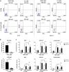
Expression profile and functional activity of CD158ab/KIR2DL-1,-2/3+ NK-cells. (a) Expression profile of selected NK-cell receptors and activation markers on CD158ab+ CD3-CD56+ NK-cells. The frequencies of CD158ab+ CD3-CD56+ NK-cells co-expressing the natural cytotoxic receptor CD335/NKp46, the activating NK receptors CD226/DNAM-1 and CD159c/NKG2C, CD16/Fc γ RIII, CD25/IL2Rα and CD279/PD-1 in peripheral blood and tumor cell suspensions of GBM patients and control individuals were determined. (b) IFN-γ and perforin expression of CD3-CD56+ NK-cells and CD158ab+ NK-cell subsets (n = 10): The proportion of IFN-γ and perforin producing cells within the entire CD3-CD56+ NK-cell population and the CD158ab+ NK-cell subsets of peripheral blood and tumor cell suspensions of GBM patients and control individuals after stimulation with PMA/Ionomycin were determined. (c) IFN-γ expression of CD56dim and CD56bright as well as CD158ab+ CD56dim and CD56bright NK-cell subsets (n = 10): The proportion of IFN-γ cells within the CD56dim and CD56bright NK-cell population and CD158ab+ CD56dim and CD56bright NK-cell subsets of peripheral blood and tumor cell suspensions of GBM patients and control individuals after stimulation with PMA/Ionomycin were determined. Mean values + /− SD are shown. P values were calculated using the Kruskal–Wallis test with post-hoc Dunn´s multiple comparison analysis (*p < 0.05,**p < 0.01, ***p < 0.001, ****p < 0.0001).
Figure 3
Figure 3
KIR2DL-1,-2/3 blockade reverses the functional hypoactivity of GBM-derived NK-cells. (a) Functional analysis of blood- and GBM-derived NK-cells: NK-cell responsiveness was assessed by cell surface mobilization of CD107a and IFN-γ production following K562 stimulation of resting or IL-2 preactivated PBMC and TIL at an effector to target ratio of 10:1 for 4.5 h. Representative dot plots are shown illustrating the capacity of blood and tumor-derived CD3-CD56+ NK-cells to express CD107a, IFN-γ or both CD107a and IFN-γ after stimulation with K562-0 cells or culture medium alone (none). (b,c) The activity and cytolytic potential of glioblastoma-derived NK-cells after KIR2DL-1,-2/3 blockade were assessed. (b) Blocking experiments revealed that Lirilumab significantly inhibited the binding of CD158a and CD158b mAb to blood- and tumor-derived NK-cells at a concentration of 30 μg/ml (****p < 0.0001). (c) IL-2 preactivated PBMC and TIL from different glioblastoma patients (n = 5) were pre-incubated with a human IgG4 isotype control mAb or Lirilumab (30 μg/ml), cocultured with culture medium (none), IFN-γ pretreated HLA-class I deficient K562 cells (K562-0) and HLA-C expressing K562 cells (K562-HLA-C) at an effector to target ratio of 10:1 for 4.5 h and then analyzed by flow cytometry. The frequencies of CD107a+, IFN-γ+and CD107a+IFN-γ+CD3-CD56+ NK-cells were determined. Mean values + /− SD are depicted. P values were calculated using the two-tailed Mann–Whitney test (*p < 0.05, **p < 0.01).
Figure 4
Figure 4
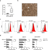
KIR2DL-1,-2/3 blockade improves the NK-cell stimulatory properties of HLA class-I expressing GBM cells. (a) Primary GBM cell cultures were prepared and evaluated for the expression of HLA class-I molecules (HLA-ABC) by flow cytometry (n = 5). (b) Representative immunohistochemical staining of HLA class I-expressing cells within glioblastoma tissue. (c) Expression of HLA class-I molecules on unstimulated (grey histograms) or IFN-γ pretreated (red histograms) K562-HLA-C and primary GBM cells (GBM1-3). (d) IL-2 preactivated PBMC from glioblastoma patients (n = 3) were pre-incubated with a human IgG4 isotype control mAb or Lirilumab (30 μg/ml), cocultured with IFN-γ pretreated HLA-C expressing K562 cells (K562-HLA-C) or primary GBM cells at an effector to target ratio of 10:1 for 4.5 h and then analyzed by flow cytometry. The frequencies of CD107a+, IFN-γ+ and CD107a+IFN-γ+ CD3-CD56+ NK-cells were determined. Mean values + /− SD are depicted. P values were calculated using the two-tailed Mann–Whitney test (*p < 0.05, **p < 0.01, ***p < 0.001, ****p < 0.0001).
Figure 5
Figure 5
Functional analysis of GBM-derived CD4+ and CD8+ memory T-cells. (a) The frequency of CD4+ and CD8+ CD45RA- memory T-cells in PBMC and TIL of GBM patients were determined by flow cytometry (n = 10). Mean values + /− SD are depicted. P values were calculated using the two-tailed Mann–Whitney test (****p < 0.0001). (b) IL-2 and (c) IFN-γ production of CD4+ and CD8+ CD45RA- memory T-cells after stimulation with culture medium (none), immobilized anti-CD3 mAb, immobilized anti-CD3/anti-CD28 mAb or PMA/Ionomycin for 6 h in the presence of Brefeldin A and Monsenin (n = 10). Mean values + /− SD from five different GBM patients are depicted. P values were calculated using the Kruskal–Wallis test with post-hoc Dunn´s multiple comparison analysis (**p < 0.01, ***p < 0.001, ****p < 0.0001).

Similar articles

Cited by

References

    1. Moretta L, Montaldo E, Vacca P, Del Zotto G, Moretta F, Merli P, Locatelli F, Mingari MC. Human natural killer cells: origin, receptors, function, and clinical applications. Int. Arch. Allergy. Immunol. 2014;164:253–264. doi: 10.1159/000365632. - DOI - PubMed
    1. Gras Navarro A, Bjorklund AT, Chekenya M. Therapeutic potential and challenges of natural killer cells in treatment of solid tumors. Front. Immunol. 2015;6:202. doi: 10.3389/fimmu.2015.00202. - DOI - PMC - PubMed
    1. Del Zotto G, Marcenaro E, Vacca P, Sivori S, Pende D, Della Chiesa M, Moretta F, Ingegnere T, Mingari MC, Moretta A, Moretta L. Markers and function of human NK cells in normal and pathological conditions. Cytometry B Clin. Cytom. 2017;92:100–114. doi: 10.1002/cyto.b.21508. - DOI - PubMed
    1. Moretta L, Moretta A. Unravelling natural killer cell function: triggering and inhibitory human NK receptors. EMBO J. 2004;23:255–259. doi: 10.1038/sj.emboj.7600019. - DOI - PMC - PubMed
    1. Sivori S, Vacca P, Del Zotto G, Munari E, Mingari MC, Moretta L. Human NK cells: surface receptors, inhibitory checkpoints, and translational applications. Cell Mol. Immunol. 2019;16:430–441. doi: 10.1038/s41423-019-0206-4. - DOI - PMC - PubMed

Publication types