. 2022 Apr 26;12(1):6805.
doi: 10.1038/s41598-022-10753-4.

Glioma facilitates the epileptic and tumor-suppressive gene expressions in the surrounding region

Affiliations

Glioma facilitates the epileptic and tumor-suppressive gene expressions in the surrounding region

Kazuki Komiyama et al. Sci Rep. .

Abstract

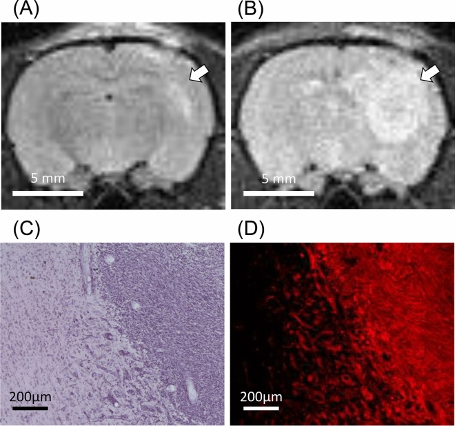
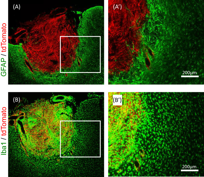
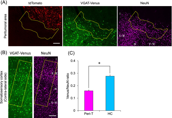
Patients with glioma often demonstrate epilepsy. We previously found burst discharges in the peritumoral area in patients with malignant brain tumors during biopsy. Therefore, we hypothesized that the peritumoral area may possess an epileptic focus and that biological alterations in the peritumoral area may cause epileptic symptoms in patients with glioma. To test our hypothesis, we developed a rat model of glioma and characterized it at the cellular and molecular levels. We first labeled rat C6 glioma cells with tdTomato, a red fluorescent protein (C6-tdTomato), and implanted them into the somatosensory cortex of VGAT-Venus rats, which specifically expressed Venus, a yellow fluorescent protein in GABAergic neurons. We observed that the density of GABAergic neurons was significantly decreased in the peritumoral area of rats with glioma compared with the contralateral healthy side. By using a combination technique of laser capture microdissection and RNA sequencing (LCM-seq) of paraformaldehyde-fixed brain sections, we demonstrated that 19 genes were differentially expressed in the peritumoral area and that five of them were associated with epilepsy and neurodevelopmental disorders. In addition, the canonical pathways actively altered in the peritumoral area were predicted to cause a reduction in GABAergic neurons. These results suggest that biological alterations in the peritumoral area may be a cause of glioma-related epilepsy.


Conflict of interest statement

The authors declare no competing interests.

Figures

Figure 1
Figure 1
Characterization of C6-tdTomato cells. (A) Phase-contrast and fluorescence microscopy images of C6-tdTomato cells. (B) Viability of C6-tdTomato cells and their parent C6 glioma cells. Cell viability was assessed by the MTT assay and repeated four times. The averaged values of OD570 with SE are demonstrated as a line graph.
Figure 2
Figure 2
Development of the rat glioma model by implantation of C6-tdTomato cells into the brain. (A,B) T2-weighted MR images of the rat brain implanted with C6-tdTomato cells at 4 days and 10 days after the surgery. The tumor area was indicated by a white arrow. (C,D) Serial sections of C6-tdTomato-implanted rat brains were made 10 days after surgery, one section was stained by Hematoxylin–Eosin staining, and another section was observed by the fluorescence microscopy.
Figure 3
Figure 3
Localization of astrocytes and microglia in the peritumoral area of C6-tdTomato implanted rats. (A,A′) Astrocytes around tumors demonstrate activated forms and highly express GFAP. (B,B′) Microglia around tumors demonstrate activated forms and highly express Iba1.
Figure 4
Figure 4
Densities of excitatory and inhibitory neurons in the peritumoral area. Representative images showing the cells labeled with tdTomato, Venus, and NeuN in the peritumoral area (A) and in the contralateral side (B) of the somatosensory cortex of the same rat. The number of Venus-positive and NeuN-positive neurons was counted in layers II to VI within the dotted line and the ratio of Venus-positive/NeuN-positive neurons was calculated (C). The data were obtained from five sections from five rats (total 25 sections). The means with SE are demonstrated as column. *P < 0.05 between healthy control (HC) and peritumoral tissues (Peri-T) (Student’s t-test). The white bars indicate 200 μm.
Figure 5
Figure 5
Representative images of brain sections before (A) and after (B) LCM.
Figure 6
Figure 6
Association image of 19 differentially expressed genes (DEGs) with diseases and functions. Ingenuity Pathway Analysis revealed that the 5 DEGs were associated with epilepsy and neurodevelopmental disorders.
Figure 7
Figure 7
Canonical pathways predicted to be actively altered in the peritumoral area. The results of canonical pathways between two groups were used as the dataset for the comparison analysis. Z-scores greater than 2 (or lower than -2) were considered significant. Significant canonical pathways specific to peritumoral tissues (Peri-T) are shown in the bar graph.

Similar articles

Cited by

References

    1. Boujelben A, et al. Multimodality imaging and mathematical modelling of drug delivery to glioblastomas. Interface Focus. 2016;6:20160039. doi: 10.1098/rsfs.2016.0039. - DOI - PMC - PubMed
    1. Stupp R, et al. Radiotherapy plus concomitant and adjuvant temozolomide for glioblastoma. N. Engl. J. Med. 2005;352:987–996. doi: 10.1056/NEJMoa043330. - DOI - PubMed
    1. Stupp R, et al. Promising survival for patients with newly diagnosed glioblastoma multiforme treated with concomitant radiation plus temozolomide followed by adjuvant temozolomide. J. Clin. Oncol. 2002;20:1375–1382. doi: 10.1200/JCO.2002.20.5.1375. - DOI - PubMed
    1. Hildebrand J, Lecaille C, Perennes J, Delattre JY. Epileptic seizures during follow-up of patients treated for primary brain tumors. Neurology. 2005;65:212–215. doi: 10.1212/01.wnl.0000168903.09277.8f. - DOI - PubMed
    1. Kurzwelly D, Herrlinger U, Simion M. Seizures in patients with low-grade gliomas–incidence, pathogenesis, surgical management, and pharmacotherapy. Adv. Tech. Stand. Neurosurg. 2010;35:81–111. doi: 10.1007/978-3-211-99481-8_4. - DOI - PubMed

Publication types