. 2022 Apr 26;12(1):6800.
doi: 10.1038/s41598-022-10961-y.

Baseline predictors for good visual gains after anti-vascular endothelial growth factor therapy for myopic choroidal neovascularization

Affiliations

Baseline predictors for good visual gains after anti-vascular endothelial growth factor therapy for myopic choroidal neovascularization

Cherng-Ru Hsu et al. Sci Rep. .

Abstract

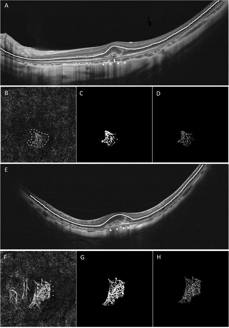
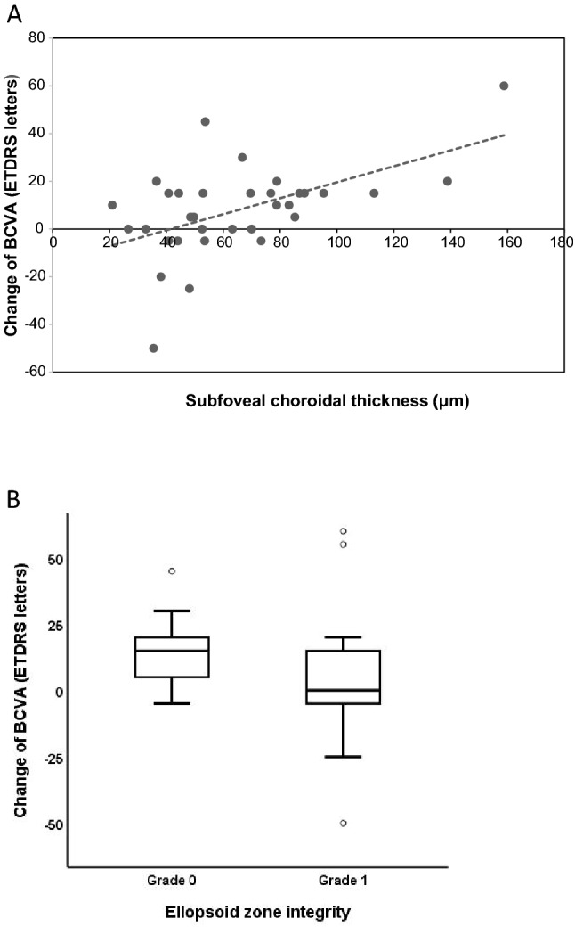
To investigate optical coherence tomography (OCT) and OCT angiography (OCTA) biomarkers for good visual outcomes in eyes with myopic choroidal neovascularization (mCNV) following anti-vascular endothelial growth factor (anti-VEGF) therapy. Patients diagnosed with mCNV via multimodal imaging were retrospectively reviewed. Baseline demographic data and biomarkers were collected. Anti-VEGF treatment based on a pro re nata (PRN) regimen was conducted on all eyes. The visual gains of ≥ 15 ETDRS letters or < 15 letters at 12-month were classified into two groups. Regression analysis was used to identify variables associated with significant best-corrected visual acuity (BCVA) improvement. Among 34 patients, 17 eyes and 17 eyes were classified into the two groups. There were no statistically significant differences in qualitative OCTA biomarkers between the two groups. The ≥ 15 letters group had significantly thicker subfoveal choroid thickness (SFCT) (79.97 ± 33.15 vs. 50.66 ± 18.31, P = 0.003), more ellipsoid zone integrity (58.8% vs. 23.5%, P = 0.037) and lower levels of fractal dimension (1.45 ± 0.101 vs. 1.53 ± 0.082, P = 0.031) than the < 15 letters group. SFCT and the ellipsoid zone integrity were correlated with 15 letters or more VA improvement in both univariable and multivariable analyses (P = 0.023 and P = 0.044, respectively). Thicker SFCT and integrity of the ellipsoid zone at baseline were associated with greater visual gains at 12 months. OCTA biomarkers seem to play a less important role in predicting the visual outcome of mCNV.


Conflict of interest statement

The authors declare no competing interests.

Figures

Figure 1
Figure 1
Baseline optical coherence tomography (OCT) and OCT angiography (OCTA) images of two patients with myopic choroidal neovascularization (mCNV). (A) This case is a 24-year-old female with an axial length of 29.20 mm. Baseline spectral-domain OCT B-scan shows a subfoveal choroidal thickness (SFCT) of 78.88 μm (double arrow). The choroid-scleral interface is indicated by white arrowheads. An ellipsoid zone disruption (graded 0) is found in the fovea (hollow arrow). (B) OCTA en-face image (3 × 3 mm) at baseline reveals a tangled-shaped NV with peripheral arcade/loops (*) and perilesional halo (white dashed line). The binarized and skeletonized image of the total NV lesion are showed in (C) and (D). Her baseline best-corrected visual acuity (BCVA) was 65 ETDRS letters, and improved to 85 EDTRS letters at 12 months after receiving one anti-vascular endothelial growth factor (anti-VEGF) injection. (E) This case is a 63-year-old female with axial length of 29.32 mm. Spectral-domain OCT B-scan at baseline shows an SFCT of 40.75 μm (double arrow). The choroid-scleral interface is indicated by white arrowheads. The absence of an ellipsoid zone (graded 1) is found in the fovea with haziness of the CNV border (hollow arrow). (F) OCTA en-face image (3 × 3 mm) at baseline shows a seafan-shaped NV with peripheral arcade/loops (*) and prominent branching vessels (white dashed square). The binarized and skeletonized image of the total NV lesion are showed in (G) and (H). Her BCVA was 70 ETDRS letters and 65 EDTRS letters at baseline and 12 months, respectively. She received a total of two anti-VEGF injections. The white lines in OCT B-scan represent the outer border of the outer plexiform layer, and the gray lines indicate the retinal pigment epithelium.
Figure 2
Figure 2
Analysis of the association between change in best-corrected visual acuity (BCVA) with subfoveal choroidal thickness (SFCT) and comparison change in BCVA with ellipsoid zone integrity. (A) The figure shows the linear regression and association analysis of SFCT with change in BCVA between baseline and 12 months (P = 0.002, r = 0.515). Dashed line represents the regression line. (B) Box plots of change in BCVA with ellipsoid zone integrity are shown. Quantitative analysis shows that there is significant difference in the grading of ellipsoid zone integrity (P = 0.049).

References

    1. Ohno-Matsui K, Ikuno Y, Lai TY, Cheung CMG. Diagnosis and treatment guideline for myopic choroidal neovascularization due to pathologic myopia. Prog. Retin. Eye Res. 2018;63:92–106. doi: 10.1016/j.preteyeres.2017.10.005. - DOI - PubMed
    1. El Matri L, Chebil A, Kort F. Current and emerging treatment options for myopic choroidal neovascularization. Clin. Ophthalmol. 2015;9:733. doi: 10.2147/OPTH.S49437. - DOI - PMC - PubMed
    1. Wong TY, Ohno-Matsui K, Leveziel N, Holz FG, Lai TY, Yu HG, et al. Myopic choroidal neovascularisation: Current concepts and update on clinical management. Br. J. Ophthalmol. 2015;99(3):289–296. doi: 10.1136/bjophthalmol-2014-305131. - DOI - PMC - PubMed
    1. Tufail A, Narendran N, Patel PJ, Sivaprasad S, Amoaku W, Browning AC, et al. Ranibizumab in myopic choroidal neovascularization: The 12-month results from the REPAIR study. Ophthalmology. 2013;120(9):1944–5.e1. doi: 10.1016/j.ophtha.2013.06.010. - DOI - PubMed
    1. Ikuno Y, Ohno-Matsui K, Wong TY, Korobelnik J-F, Vitti R, Li T, et al. Intravitreal aflibercept injection in patients with myopic choroidal neovascularization: The MYRROR study. Ophthalmology. 2015;7(6):1220–1227. doi: 10.1016/j.ophtha.2015.01.025. - DOI - PubMed

MeSH terms