. 2022 Apr 26;7(1):49.
doi: 10.1038/s41541-022-00461-5.

Two DNA vaccines protect against severe disease and pathology due to SARS-CoV-2 in Syrian hamsters

Affiliations

Two DNA vaccines protect against severe disease and pathology due to SARS-CoV-2 in Syrian hamsters

George Giorgi Babuadze et al. NPJ Vaccines. .

Abstract

The SARS-CoV-2 pandemic is an ongoing threat to global health, and wide-scale vaccination is an efficient method to reduce morbidity and mortality. We designed and evaluated two DNA plasmid vaccines, based on the pIDV-II system, expressing the SARS-CoV-2 spike gene, with or without an immunogenic peptide, in mice, and in a Syrian hamster model of infection. Both vaccines demonstrated robust immunogenicity in BALB/c and C57BL/6 mice. Additionally, the shedding of infectious virus and the viral burden in the lungs was reduced in immunized hamsters. Moreover, high-titers of neutralizing antibodies with activity against multiple SARS-CoV-2 variants were generated in immunized animals. Vaccination also protected animals from weight loss during infection. Additionally, both vaccines were effective at reducing both pulmonary and extrapulmonary pathology in vaccinated animals. These data show the potential of a DNA vaccine for SARS-CoV-2 and suggest further investigation in large animal and human studies could be pursued.


Conflict of interest statement

The authors declare no competing interests.

Figures

Fig. 1
Fig. 1. Design of plasmid vaccines.
a Diagram showing the pIDV-II vector and antigens. Both plasmids encode codon-optimized full-length SARS-CoV-2 spike (S) open reading frame in the presence or absence of 5mer4 at 3′ end. b Detection of SARS-CoV-2 Spike protein in HEK 293 cells lysate by western blot. Cells were transfected with pIDV-II-SARSoCoV-Spike_v1 (pIDV-V1) and pIDV-II-SARSoCoV-Spike_v5 (pIDV-V5). The mock sample represents the 293 T cells transfected with empty vector pIDV-II; Western blot was performed using a convalescent human serum.
Fig. 2
Fig. 2. Humoral and cell-mediated responses in vaccinated mice.
a BALB/c and b C57BL/6 mice were immunized with pIDV-II-SARS-CoV2-Spike-V1 (pIDV-V1), pIDV-II-SARS-CoV2-Spike-V5 (pIDV-V5), or sham vaccination with buffer only (control). Spike-specific antibodies was detected by ELISA. All mice received two doses of vaccine, one on day 0 and another on day 28 (n = 4 per group per timepoint). Each dose of vaccine was administered via electroporation (EP) following intramuscular injection of 50 µg/dose (ug DNA by IM + EP route). (sera dilution 1:400). **** indicates P value = <0.0001 as determined by one-way ANOVA. Error bars represent standard deviation. T cell response was analyzed by ELISpot 10 days after boost in c BALB/c and d C57BL/6 mice. Sham immunized mice were used as control. Splenocytes cell suspension were stimulated with SARS-CoV2 peptide pools encompassing the entire SARS-CoV2 Spike glycoprotein. No significant difference was observed for the cell-mediated response between vaccinated animals with two versions of vaccines.
Fig. 3
Fig. 3. Weight loss in animals following virus challenge.
Animals were weighed throughout the course of infection and weight change was compared to pre-infection weight (n = 8 per group). Error bars represent standard deviation. All * represent timepoints where P value <0.0001 as determined by two-way ANOVA.
Fig. 4
Fig. 4. Vaccination reduces viral shedding and viral RNA load in SARS-Cov2 challenged hamsters.
a Vaccination and challenge schedule. Sampling timepoints are shown by black arrows. Groups of animals (n = 4) were euthanized at 4 and 8 dpi as shown by the orange arrows. Sampling timepoints are shown by black arrows. Titers of infectious virus from various timepoints were collected and determined by TCID50 from b oral swabs, c nasal washes, d nasal turbinates, and e lungs. Error bars represent standard deviation. P value = 0.0001 (****) and 0.0003 (***).
Fig. 5
Fig. 5. Comparison of SARS-CoV-2 viral load in different tissues.
a Viral RNA load (mean [SD]) from hamster lung samples in control, PIDV-V1, and PIDV-V5 groups at day 4 and day 8 post-infection. Two-way- ANOVA- Dunnett’s multiple comparisons test was performed (Control vs. PIDV-V1 day 4, **p = 0.0091, Control vs. PIDV-V5 day 4 *p = 0.0457, Control vs. PIDV-V1 day 8, *p = 0.0101, Control vs. PIDV-V5 day 8 *p = 0.0119). b Viral load from hamster nasal turbinate samples in control, PIDV-V1, and PIDV-V5 groups at day 4 and day 8 post-infection. Two-way- ANOVA- Dunnett’s multiple comparisons test was performed (Control vs. PIDV-V1 day 4, p = 0.1393, Control vs. PIDV-V5 day 4 p = 0.5203, Control vs. PIDV-V1 day 8, p = 0.3616, Control vs. PIDV-V5 day 8 p = 0.4596). c Viral load from hamster Kidney samples in control, PIDV-V1, and PIDV-V5 groups at day 4 and day 8 post-infection. Two-way- ANOVA- Dunnett’s multiple comparisons test was performed (Control vs. PIDV-V1 day 4, p = 0.3591, Control vs. PIDV-V5 day 4 p = 0.3768, Control vs. PIDV-V5 day 8 p = 0.3432). For all panels, error bars represent the standard deviation.
Fig. 6
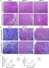
Fig. 6. Lung pathology in vaccinated and unvaccinated hamsters.
a Representative HE stain (10X magnification, Scale bar = 100 um) in PIDV-V1, PIDV-V5, and control groups at 4 and 8 days post-infection (n = 4 per group). b Representative Masson’s trichrome staining (10X magnification, Scale bar = 100 um). Collagen is indicated by areas stained in blue. c Quantification of fibrosis (collagen staining) presented as mean percentage fibrosis of total lung tissue. Two-way- ANOVA- Dunnett’s multiple comparisons test was performed (Control vs. PIDV-V1 day 4, ****p < 0.0001, Control vs. PIDV-V5 day 4 ****p < 0.0001, Control vs. PIDV-V1 day 8, ****p < 0.0001, Control vs. PIDV-V5 day 8 ****p < 0.0001). Scale bar represents 100 μm.
Fig. 7
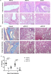
Fig. 7. Histopathology of kidneys in vaccinated and unvaccinated hamsters.
a Representative H&E staining (10X magnification, Scale bar = 100 um) in pIDV-V1, pIDV-V5, and control groups at 4, 8 days post-infection (n = 4 per group). b Representative Masson’s trichrome staining (10X magnification, Scale bar = 100 um) in pIDV-V1, pIDV-V5, and control groups at 4, 8 days post-infection (n = 4). Collagen is indicated by areas stained in blue. c Quantification of inflammatory cell infiltration in kidney tissues of control, pIDV-VI, and pIDV-V5 vaccinated groups. Two-way- ANOVA- Dunnett’s multiple comparisons test was performed (Control vs. pIDV-V1 day 4, **p = 0.0011, Control vs. pIDV-V5 day 4 **p = 0.0017, Control vs. pIDV-V1 day 8, ***p = 0.0001, Control vs. pIDV-V5 day 8 ****p = 0.0404). d Quantification of fibrosis (collagen staining) presented as mean percentage fibrosis of total kidney tissue. Two-way- ANOVA- Dunnett’s multiple comparisons test was performed (Control vs. pIDV-V1 day 4, *p = 0.0241, Control vs. pIDV-V5 day 4 **p = 0.0057, Control vs. pIDV-V1 day 8, **p = 0.0077, Control vs. pIDV-V5 day 8 *p = 0.0120). Scale bar represents 100 μm.
Fig. 8
Fig. 8. Virus neutralization.
a Serum neutralizing titers from hamsters vaccinated with TE buffer (control), pIDV-V1, pIDV-V5 were analysed against the SB3 (B1) isolate 4 weeks after receiving the second vaccination and prior to challenge. Titers were also evaluated at 4 and 8 dpi against b Alpha (B.1.1.7) variant and c Beta (B.1.351) as well as d SB3 (B1). Four hamsters were used per group. Two-way ANOVA with repeated measures was used for analysis, and stars denote p < 0.0001.

Similar articles

Cited by

References

    1. Li Q, et al. Early transmission dynamics in Wuhan, China, of novel coronavirus-infected pneumonia. N. Engl. J. Med. 2020;382:1199–1207. doi: 10.1056/NEJMoa2001316. - DOI - PMC - PubMed
    1. Zhu N, et al. A novel coronavirus from patients with pneumonia in China, 2019. N. Engl. J. Med. 2020;382:727–733. doi: 10.1056/NEJMoa2001017. - DOI - PMC - PubMed
    1. Nalbandian, A. et al. Post-acute COVID-19 syndrome. Nat. Med.10.1038/s41591-021-01283-z (2021). - PMC - PubMed
    1. Kozak, R. et al. Recognition of long-COVID-19 patients in a Canadian tertiary hospital setting: a retrospective analysis of their clinical and laboratory characteristics. Pathogens10.3390/pathogens10101246 (2021). - PMC - PubMed
    1. Polack FP, et al. Safety and efficacy of the BNT162b2 mRNA Covid-19 vaccine. N. Engl. J. Med. 2020;383:2603–2615. doi: 10.1056/NEJMoa2034577. - DOI - PMC - PubMed