Molecular and neurological features of MELAS syndrome in paediatric patients: A case series and review of the literature
- PMID: 35474314
- PMCID: PMC9266612
- DOI: 10.1002/mgg3.1955
Molecular and neurological features of MELAS syndrome in paediatric patients: A case series and review of the literature
Abstract
Background: Mitochondrial encephalomyopathy, lactic acidosis and stroke-like episodes (MELAS) syndrome is one of the most well-known mitochondrial diseases, with most cases attributed to m.3243A>G. MELAS syndrome patients typically present in the first two decades of life with a broad, multi-systemic phenotype that predominantly features neurological manifestations--stroke-like episodes. However, marked phenotypic variability has been observed among paediatric patients, creating a clinical challenge and delaying diagnoses.
Methods: A literature review of paediatric MELAS syndrome patients and a retrospective analysis in a UK tertiary paediatric neurology centre were performed.
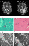
Results: Three children were included in this case series. All patients presented with seizures and had MRI changes not confined to a single vascular territory. Blood heteroplasmy varied considerably, and one patient required a muscle biopsy. Based on a literature review of 114 patients, the mean age of presentation is 8.1 years and seizures are the most prevalent manifestation of stroke-like episodes. Heteroplasmy is higher in a tissue other than blood in most cases.
Conclusion: The threshold for investigating MELAS syndrome in children with suspicious neurological symptoms should be low. If blood m.3243A>G analysis is negative, yet clinical suspicion remains high, invasive testing or further interrogation of the mitochondrial genome should be considered.
Keywords: MELAS syndrome; encephalopathy; genetics; lactic acidosis; m.3243A>G; mitochondrial disease; paediatric neurology; stroke-like episodes.
© 2022 The Authors. Molecular Genetics & Genomic Medicine published by Wiley Periodicals LLC.
Conflict of interest statement
The authors declare no potential conflict of interest.
Figures


References
-
- Alcubilla‐Prats, P. , Solé, M. , Botey, A. , Grau, J. M. , Garrabou, G. , & Poch, E. (2017). Kidney involvement in MELAS syndrome: Description of 2 cases. Medicina Clínica (Barcelona), 148, 357–361. - PubMed
-
- Anan, R. , Nakagawa, M. , Miyata, M. , Higuchi, I. , Nakao, S. , Suehara, M. , Osame, M. , & Tanaka, H. (1995). Cardiac involvement in mitochondrial diseases: A study on 17 patients with documented mitochondrial DNA defects. Circulation, 91, 955–961. - PubMed
-
- Brisca, G. , Fiorillo, C. , Nesti, C. , Trucco, F. , Derchi, M. , Andaloro, A. , Assereto, S. , Morcaldi, G. , Pedemonte, M. , Minetti, C. , Santorelli, F. M. , & Bruno, C. (2015). Early onset cardiomyopathy associated with the mitochondrial tRNALeu(UUR) 3271T>C MELAS mutation. Biochemical and Biophysical Research Communications, 458, 601–604. - PubMed
-
- Calvaruso, M. A. , Willemsen, M. A. , Rodenburg, R. J. , van den Brand, M. , Smeitink, J. A. M. , & Nijtmans, L. (2011). New mitochondrial tRNAHIS mutation in a family with lactic acidosis and stroke‐like episodes (MELAS). Mitochondrion, 11, 778–782. - PubMed
Publication types
MeSH terms
Grants and funding
LinkOut - more resources
Full Text Sources
Medical

