Review
. 2022 Apr;54(4):377-392.
doi: 10.1038/s12276-021-00677-w. Epub 2022 Apr 26.

Role of the gut-brain axis in energy and glucose metabolism

Affiliations
Review

Role of the gut-brain axis in energy and glucose metabolism

Hallie R Wachsmuth et al. Exp Mol Med. 2022 Apr.

Abstract

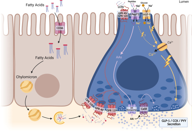
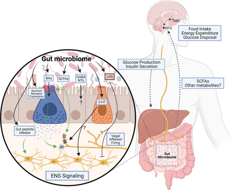
The gastrointestinal tract plays a role in the development and treatment of metabolic diseases. During a meal, the gut provides crucial information to the brain regarding incoming nutrients to allow proper maintenance of energy and glucose homeostasis. This gut-brain communication is regulated by various peptides or hormones that are secreted from the gut in response to nutrients; these signaling molecules can enter the circulation and act directly on the brain, or they can act indirectly via paracrine action on local vagal and spinal afferent neurons that innervate the gut. In addition, the enteric nervous system can act as a relay from the gut to the brain. The current review will outline the different gut-brain signaling mechanisms that contribute to metabolic homeostasis, highlighting the recent advances in understanding these complex hormonal and neural pathways. Furthermore, the impact of the gut microbiota on various components of the gut-brain axis that regulates energy and glucose homeostasis will be discussed. A better understanding of the gut-brain axis and its complex relationship with the gut microbiome is crucial for the development of successful pharmacological therapies to combat obesity and diabetes.


Conflict of interest statement

The authors declare no competing interests.

Figures

Fig. 1
Fig. 1. Major mediators of the gut–brain axis.
Specialized intestinal epithelial cells, enteroendocrine cells (EECs), neuropod cells, and enterochromaffin cells (ECs), secrete gut peptides, including GLP-1, CCK, GIP, and PYY, on the basolateral side. These gut peptides are released in close proximity to vagal afferent neurons innervating the intestinal mucosa and activate these neurons. Vagal afferent neurons send signals to the nucleus tractus solitarius (NTS), which can send signals to higher-order brain regions, such as the arcuate nucleus (ARC). Vagal afferent neurons are also activated via the enteric nervous system, which can be activated by the release of gut-derived neurotransmitters, such as 5-HT, from ECs and intraganglionic laminar endings (IGLEs) sensing intestinal stretch. In addition, gut peptides are able to enter the circulation and carry signals directly to the NTS or activate splanchnic nerve endings to signal to the brain via spinal afferent neurons.
Fig. 2
Fig. 2. Nutrient sensing by enteroendocrine cells.
The different macronutrients act through alternate pathways to elicit gut peptide release. Fatty acids can signal through multiple receptors on both the apical and basolateral membranes. Signaling at the basolateral membrane requires the uptake and packaging of fats into chylomicrons in enterocytes followed by the release and breakdown of these chylomicrons at the basolateral surface. Fatty acids bind their receptors on enteroendocrine cells, which activate a downstream signaling cascade leading to the fusion of gut peptide–containing vesicles and the release of their contents at the basolateral membrane. Glucose sensing occurs at the apical membrane of an EEC and requires uptake into the cell, along with Na+, via SGLT-1. Na+ entry into the EEC causes depolarization and subsequent activation of Ca2+ channels, resulting in vesicle fusion and gut peptide release. Amino acid signaling in the enteroendocrine cell involves the uptake of peptides and Na+ via PepT1 at the apical membrane. This Na+ may depolarize the cells, but research is still needed to determine its exact mechanism of action. Once a peptide enters the cell, it is broken down into amino acids, which are transported out of the cell through the basolateral membrane, where they can activate CaSR, leading to Ca2+ release and vesicle fusion. CaSR may also be present at the apical membrane, but research is still needed to elucidate the exact mechanism of protein-induced gut peptide release.
Fig. 3
Fig. 3. Impact of the microbiome on gut–brain signaling.
The microbiota produces several metabolites that impact gut–brain signaling directly and indirectly. The microbiome can directly influence nutrient receptor expression and gut peptide release from EECs and can influence the production of serotonin (5-HT) from ECs, which may alter ENS or vagal afferent activation. Microbially derived or modified metabolites also influence the gut–brain axis. Bile acids (BAs) can alter the expression of TGR5 and regulate gut peptide release from EECs, while SCFAs alter nutrient receptor expression and gut peptide production by EECs or activate vagal afferents directly. Alternatively, metabolites such as LPS, a product of pathogenic microbes, can impair gut–brain signaling by preventing the activation of vagal afferents or the ENS. Overall, the gut microbiome and its metabolites can alter gut–brain signaling to influence the activation of the NTS and ARC, which regulate food intake, energy expenditure, glucose disposal and production, and insulin secretion.

References

    1. Hales, C. M., Carroll, M. D., Fryar, C. D. & Ogden, C. L. Prevalence of obesity and severe obesity among adults: United States, 2017–2018. NCHS Data Brief, 1–8 (2020). - PubMed
    1. Saeedi P, et al. Global and regional diabetes prevalence estimates for 2019 and projections for 2030 and 2045: results from the International Diabetes Federation Diabetes Atlas, 9(th) edition. Diabetes Res. Clin. Pract. 2019;157:107843. doi: 10.1016/j.diabres.2019.107843. - DOI - PubMed
    1. Leung MYM, Carlsson NP, Colditz GA, Chang S-H. The burden of obesity on diabetes in the United States: medical expenditure panel survey, 2008 to 2012. Value Health. 2017;20:77–84. doi: 10.1016/j.jval.2016.08.735. - DOI - PMC - PubMed
    1. Sjöström L. Bariatric surgery and reduction in morbidity and mortality: experiences from the SOS study. Int. J. Obes. 2008;32:S93–S97. doi: 10.1038/ijo.2008.244. - DOI - PubMed
    1. Reid T. Choosing GLP-1 receptor agonists or DPP-4 inhibitors: weighing the clinical trial evidence. Clin. Diabetes. 2012;30:3. doi: 10.2337/diaclin.30.1.3. - DOI

Publication types