. 2022 Jul;59(7):4044-4064.
doi: 10.1007/s12035-022-02818-2. Epub 2022 Apr 27.

Involvement of the Voltage-Gated Calcium Channels L- P/Q- and N-Types in Synapse Elimination During Neuromuscular Junction Development

Affiliations

Involvement of the Voltage-Gated Calcium Channels L- P/Q- and N-Types in Synapse Elimination During Neuromuscular Junction Development

Neus Garcia et al. Mol Neurobiol. 2022 Jul.

Abstract

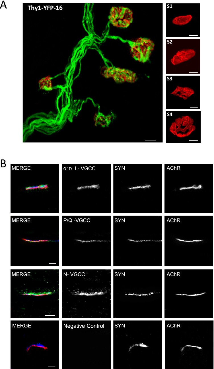
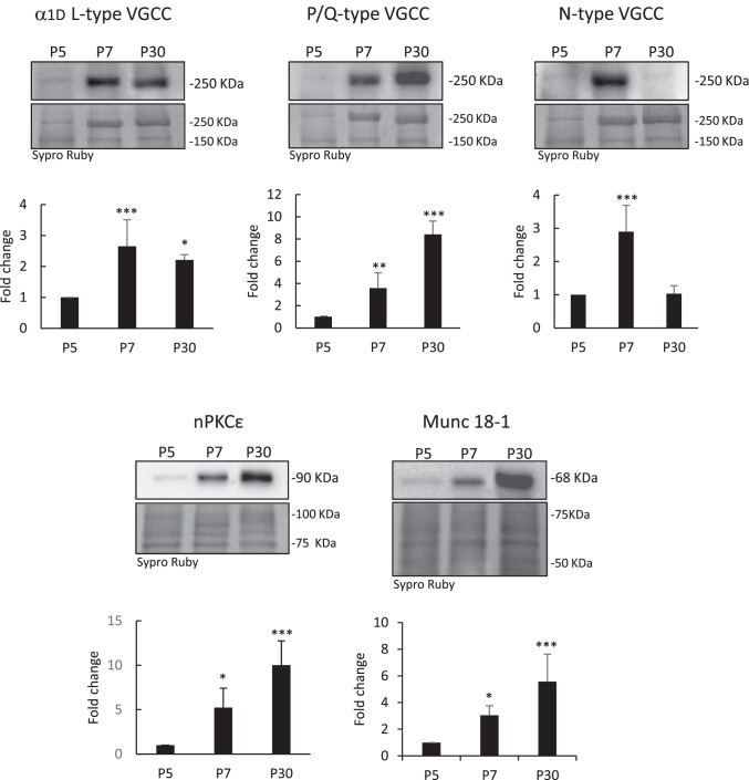
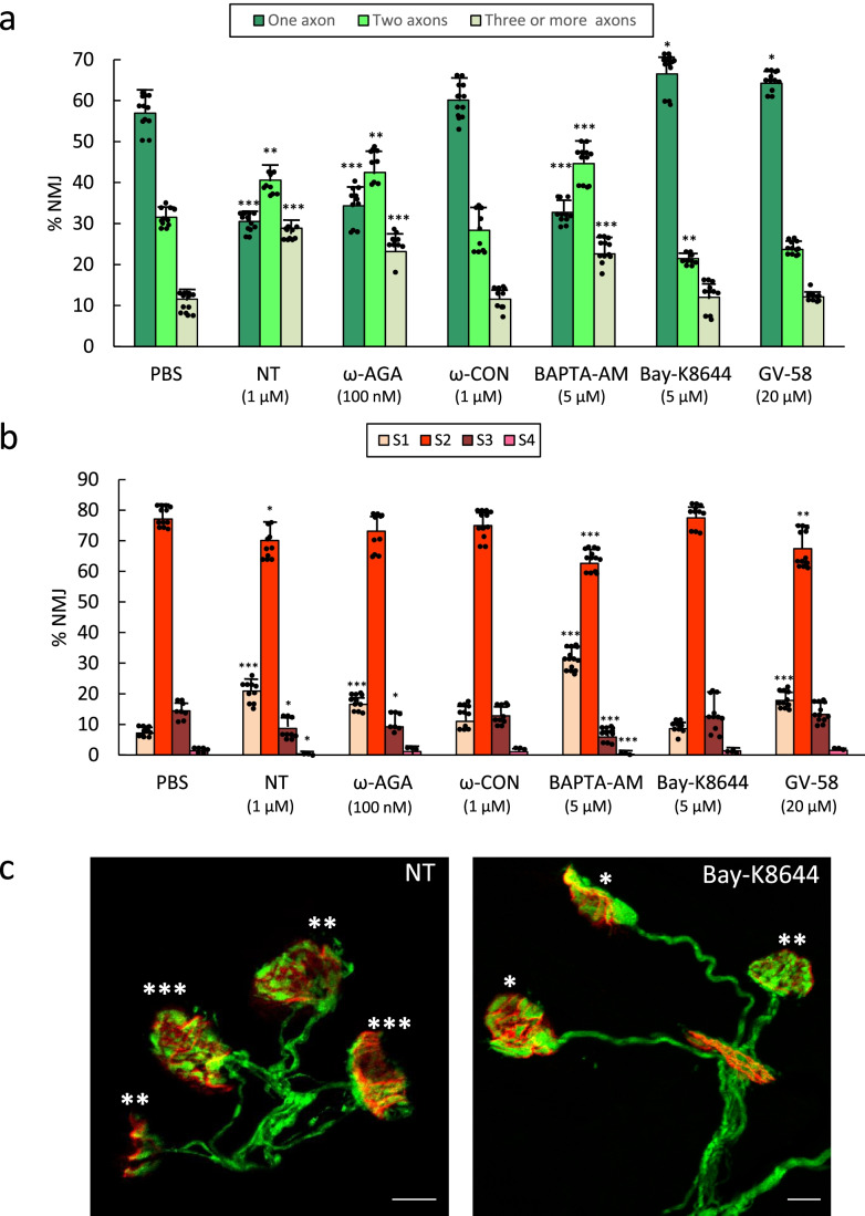
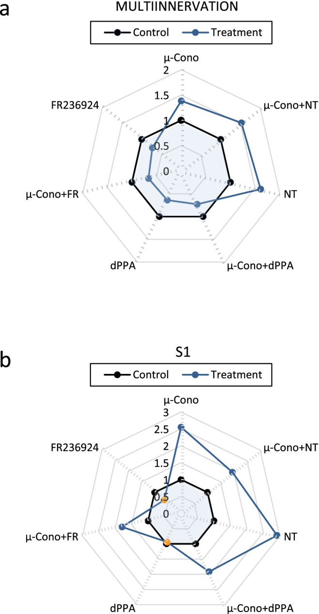
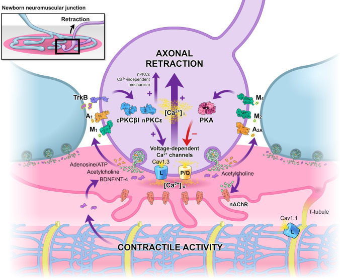
During the nervous system development, synapses are initially overproduced. In the neuromuscular junction (NMJ) however, competition between several motor nerve terminals and the synapses they made ends with the maturation of only one axon. The competitive signaling between axons is mediated by the differential activity-dependent release of the neurotransmitter ACh, co-transmitters, and neurotrophic factors. A multiple metabotropic receptor-driven downstream balance between PKA and PKC isoforms modulates the phosphorylation of targets involved in transmitter release and nerve terminal stability. Previously, we observed in the weakest endings on the polyinnervated NMJ that M1 mAChR receptors reduce ACh release through the PKC pathway coupled to an excess of Ca2+ inflow through P/Q- N- and L-type voltage-gated calcium channels (VGCC). This signaling would contribute to the elimination of this nerve terminal. Here, we investigate the involvement of the P/Q-, N-, and L-subtype channels in transgenic B6.Cg-Tg (Thy1-YFP)16-Jrs/J mice during synapse elimination. Then, the axon number and postsynaptic receptor cluster morphologic maturation were evaluated. The results show that both L- and P/Q-type VGCC (but not the N-type) are equally involved in synapse elimination. Their normal function favors supernumerary axonal loss by jointly enhancing intracellular calcium [Ca2+]i. The block of these VGCCs or [Ca2+]i i sequestration results in the same delay of axonal loss as the cPKCβI and nPKCε isoform block or PKA activation. The specific block of the muscle cell's contraction with μ-conotoxin GIIIB also delays synapse maturation, and thus, a retrograde influence from the postsynaptic site regulating the presynaptic CaV1.3 may contribute to the synapse elimination.

Keywords: Axonal competition; Motor endplate; Postnatal synapse elimination; Protein kinases; VGCC.


Conflict of interest statement

The authors declare no competing interests.

Figures

Fig. 1
Fig. 1
a Representative confocal image of a nerve terminal arborization. Singly, dually, and innervated by three or more axons NMJs from YFP muscles and also images of the morphologic maturation (S1, the most inmature, and S4, almost fully differentiated, stages) of the postsynaptic clusters from P9 mice. The bar indicates 10 μm. b Confocal immunofluorescence location of α1D L-, N-, and P/Q-type voltage-dependent calcium channels (VDCCs) at the NMJ. Triple labeling of VDCCs (green fluorescence) with syntaxin (blue fluorescence) and nAChR-α-bungarotoxin (red fluorescence) in merge images. Figure shows the presence of α1D L-, N-, and P/Q-type-VDCC (in green) in the nerve terminal of P9 Levator auris longus (LAL) muscle endplates. The bar indicates 10 μm
Fig. 2
Fig. 2
Western blots and histograms of α1D-L-, N-, and P/Q VGCC proteins in the LAL muscle of mice during development (P5-P7-P30). The developmental change in the P/Q VGCC protein level is parallel with the changes observed in other presynaptic molecules (nPKCε isoform and Munc18-1). Data are mean value ± SD, *p < 0.05, **p < 0.01, ***p < 0.005 (n = 5; 3 repeats)
Fig. 3
Fig. 3
In (a) we show the percentage of singly- and polyinnervated NMJ after 4 applications over the LAL surface (one application every day between P5–P8 (observation at P9) of one of the following VGCC inhibitor substances: nitrendipine (NT 1 μM, an L-type channel blocker), ω-conotoxin-GVIA (ω-CON 1 μM, N-type channel blocker), and ω-agatoxin-IVA (ω-AGA 100 nM, P/Q-type blocker). Also, the L activator Bay-K8644 (5 μM), the P/Q- and N-type activator GV-58 (20 μM), and the intracellular calcium chelator BAPTA-AM (5 μM). The histogram in (b) shows the percentage of S1-S4 clusters in the untreated control mice (PBS) and after the 4 applications of the aforesaid substances. Data were presented as percentages of NMJ ± SD. Fisher’s test: * p < 0.05, ** p < 0.01, *** p < 0.005. The confocal images in (c) show examples of representative NMJ areas with singly, dually, and innervated by three or more axons (the corresponding number of asterisks) from YFP muscles. At the left, the L-type channel blocker nitrendipine (NT) delays axon loss because many multi-innervated NMJs persist. By the contrary, at the right, the L activator Bay-K8644 increases the number of monoinnervated junctions. The bar indicates 10 μm
Fig. 4
Fig. 4
Diagrams are graphic representations that collectively show the pattern of action of the different agents and combinations of agents used. a Multi-innervation values. b Postsynaptic cluster (S1) values. The line between the blue and white areas means the ratio 1 (experimental value/control value) or “no effect.” Orange filled circles mean that the experimental value is not significantly different from the control value (p < 0.05). and filled circles that there is a significant difference (p > 0.05). SEMs are eliminated for clarity
Fig. 5
Fig. 5
Graphic representation of the results. The activity-dependent signaling between the nerve terminals that are in competition through several metabotropic receptors can result in the modulation of the downstream effector kinases, specifically cPKCβI, nPKCε, and PKA. Changes in kinases activity can allow the coordinate phosphorylation of the L-type CaV1.3 and P/Q-type VGCC. The high calcium entry through these operative channels present in immature nerve endings can result in their final loss. Also, muscle CaV1.1 and contractile activity can contribute to the synapse elimination. A component of this mechanism may be mediated by a retrograde influence from the postsynaptic site, via the BDNF-TrkB pathway, on the presynaptic calcium channels

Similar articles

Cited by

References

    1. Benoit P, Changeux JP. Consequences of tenotomy on the evolution of multiinnervation in developing rat soleus muscle. Brain Res. 1975;99:354–358. doi: 10.1016/0006-8993(75)90036-0. - DOI - PubMed
    1. Lichtman JW. The reorganization of synaptic connexions in the rat submandibular ganglion during post-natal development. J Physiol. 1977;273:155–177. doi: 10.1113/jphysiol.1977.sp012087. - DOI - PMC - PubMed
    1. Lanuza MA, Santafé MM, Garcia N, et al. Protein kinase C isoforms at the neuromuscular junction: Localization and specific roles in neurotransmission and development. J Anat. 2014;224:61–73. doi: 10.1111/joa.12106. - DOI - PMC - PubMed
    1. Nadal L, Garcia N, Hurtado E et al (2016) Presynaptic muscarinic acetylcholine autoreceptors (M1, M2 and M4 subtypes), adenosine receptors (A1 and A2A) and tropomyosin-related kinase B receptor (TrkB) modulate the developmental synapse elimination process at the neuromuscular junction. Mol Brain 9. 10.1186/s13041-016-0248-9 - PMC - PubMed
    1. Tomàs J, Garcia N, Lanuza MA, et al (2017) Presynaptic membrane receptors modulate ACh release, axonal competition and synapse elimination during neuromuscular junction development.Front Mol Neurosci 10(132):1–12. 10.3389/FNMOL.2017.00132 - PMC - PubMed