. 2022 Apr 6;7(15):12624-12636.
doi: 10.1021/acsomega.1c06677. eCollection 2022 Apr 19.

Spontaneous Coassembly of the Protein Terthiophene into Fluorescent Electroactive Microfibers in 2D and 3D Cell Cultures

Affiliations

Spontaneous Coassembly of the Protein Terthiophene into Fluorescent Electroactive Microfibers in 2D and 3D Cell Cultures

Ilaria Elena Palamà et al. ACS Omega. .

Abstract

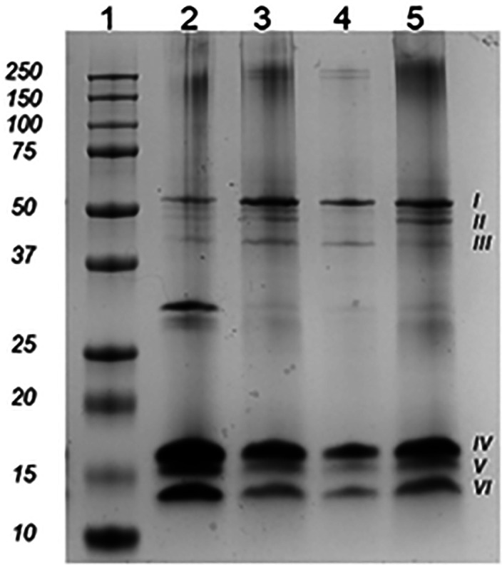
Protein-based microfibers are biomaterials of paramount importance in materials science, nanotechnology, and medicine. Here we describe the spontaneous in situ formation and secretion of nanostructured protein microfibers in 2D and 3D cell cultures of 3T3 fibroblasts and B104 neuroblastoma cells upon treatment with a micromolar solution of either unmodified terthiophene or terthiophene modified by mono-oxygenation (thiophene → thiophene S-oxide) or dioxygenation (thiophene → thiophene S,S-dioxide) of the inner ring. We demonstrate via metabolic cytotoxicity tests that modification to the S-oxide leads to a severe drop in cell viability. By contrast, unmodified terthiophene and the respective S,S-dioxide cause no harm to the cells and lead to the formation and secretion of fluorescent and electroactive protein-fluorophore coassembled microfibers with a large aspect ratio, a micrometer-sized length and width, and a nanometer-sized thickness, as monitored in real-time by laser scanning confocal microscopy (LSCM). With respect to the microfibers formed by unmodified terthiophene, those formed by the S,S-dioxide display markedly red-shifted fluorescence and an increased n-type character of the material, as shown by macroscopic Kelvin probe in agreement with cyclovoltammetry data. Electrophoretic analyses and Q-TOF mass spectrometry of the isolated microfibers indicate that in all cases the prevalent proteins present are vimentin and histone H4, thus revealing the capability of these fluorophores to selectively coassemble with these proteins. Finally, DFT calculations help to illuminate the fluorophore-fluorophore intermolecular interactions contributing to the formation of the microfibers.


Conflict of interest statement

The authors declare no competing financial interest.

Figures

Scheme 1
Scheme 1. Structures of Unmodified Terthiophene (1) and Terthiophene Modified by Mono-Oxygenation (2) or Dioxygenation (3) of the Inner Ring
Scheme 2
Scheme 2. Synthetic Pattern for the Preparation of 13
Reagents and conditions are as follows: (i) 2-thienylthiophene, [1,1′-bis(diphenylphosphino)ferrocene]dichloropalladium(II), Na2CO3 and THF/H2O (2:1) at 80 °C for 25 min. MW = microwave.
Figure 1
Figure 1
(A) Normalized absorption and photoluminescence spectra of compounds 13 in CH2Cl2. The excitation wavelength is at the maximum absorption. (B) Cyclic voltammograms recorded in 0.2 mmol L–1 (C4H9)4NClO4 in CH2Cl2 and (C) HOMO–LUMO energy diagram of compounds 13.
Figure 2
Figure 2
MTT cytotoxicity tests on (A) 3T3 and (B) B104 cells treated with compounds 13 compared to untreated cells (CTR). Representative measurements were taken from three distinct sets of data, and no significant difference between values at different time points was observed at P < 0.05 with Student’s t-test.
Figure 3
Figure 3
LSCM images of live 3T3 fibroblasts upon the uptake of (A) 1 and (B) 3 in a time window from 1 h to 7 days. Scale bars represent 25 μm. The insets show magnified details of the fiber’s elongation and thickening steps.
Figure 4
Figure 4
LSCM images of live mouse B104 neuroblastoma cells upon the uptake of (A) 1 and (B) 3 in a time window from 1 h to 7 days. The insets show the lateral assembly of several fibrils or the formation of bundles of microfibers. Scale bars represent 36 μm.
Figure 5
Figure 5
z-Stack sections acquired from photoluminescence reconstruction in the z-direction of the qualitative fiber production in (A and C) 3D fibroblasts (3T3) and (B and D) neuroblastoma cells (B104) after eight days of incubation with dyes 1 and 3, respectively. Penetration efficacy analysis of dyes (E) 1 and (F) 3 assessed by fluorescence flow cytometry after eight days in 3D spheroids. Scale bars represent 50 μm. A representative result of three independent experiments is shown.
Figure 6
Figure 6
Representative SDS-PAGE of isolated fibers produced by 3T3 fibroblasts (lanes 2 and 3) and B104 neuroblastoma cells (lanes 4 and 5) after incubation with 1 (lanes 2 and 4) and 3 (lanes 3 and 5). Lane 1 shows proteins (markers) of known molecular mass (sizes in kilodaltons are shown on the left). The most representative protein bands identified are named from I to VI (on the right).
Figure 7
Figure 7
LSCM images of the colocalization experiment between isolated green fluorescent microfibers secreted by (A) 3T3 fibroblasts and (B) B104 mouse neuroblastoma cells treated with 1 and an antivimentin antibody (red). Scale bars represent (A) 10 and (B) 5 μm. Similarly, LSCM images of the colocalization experiment between isolated red fluorescent microfibers secreted by (C) 3T3 fibroblasts and (D) B104 mouse neuroblastoma cells upon treatment with 3 and antivimentin antibody (green). Scale bars represent 10 μm.
Figure 8
Figure 8
AFM images of isolated microfibers formed by 3T3 fibroblasts upon the uptake of (A) 1 and (B) 3.Panels A1 and B1 are 3D reconstructions of the corresponding fibers in panels A and B.
Figure 9
Figure 9
(A and B) Work function values (the difference between the Fermi level and the vacuum energy) of red and green fibers obtained from 3T3 cells upon treatment with 1 or 3 measured by a macroscopic Kelvin probe. Red fibers have a lower work function absolute value (EF closer to vacuum), which is compatible with an increase of the n-type character with respect to green fibers as exemplified in the scheme in panel B.
Figure 10
Figure 10
(A) DFT-calculated structures of the dimer of fluorophores 2 (left) and 3 (right). The green region between the two molecules is the noncovalent interaction (NCI) indicator isosurface identifying the nonbonding interaction region between the two molecules. (B) A plot reporting the values of the NCI indicator for the dimers of 1, 2, and 3 (from top down). An interaction is present for the reduced density gradient (RDG) tending to zero. If this occurs for small and negative values of sign(λ2)ρ, the interaction is van der Waals-type. For larger values, the interaction is electrostatic.

References

    1. DeFrates K. G.; Moore R.; Borgesi J.; Lin G.; Mulderig T.; Beachley V.; Hu X. Protein-Based Fiber Materials in Medicine: A Review. Nanomaterials 2018, 8 (7), 457.10.3390/nano8070457. - DOI - PMC - PubMed
    2. From NLM.

    1. Wang M.-D.; Huang Y.-Q.; Wang H.. In Vivo Self-Assembly of Polypeptide-Based Nanomaterials. In Handbook of Macrocyclic Supramolecular Assembly; Liu Y., Chen Y., Zhang H.-Y., Eds.; Springer Singapore, 2019; pp 1023–1043.
    1. Schreiber S. L. Chemical genetics resulting from a passion for synthetic organic chemistry. Bioorg. Med. Chem. 1998, 6 (8), 1127–1152. 10.1016/S0968-0896(98)00126-6. - DOI - PubMed
    2. From NLM.

    3. Schreiber S. L. A Chemical Biology View of Bioactive Small Molecules and a Binder-Based Approach to Connect Biology to Precision Medicines. Isr. J. Chem. 2019, 59 (1–2), 52–59. 10.1002/ijch.201800113. - DOI - PMC - PubMed
    1. Cheng P. N.; Liu C.; Zhao M.; Eisenberg D.; Nowick J. S. Amyloid β-sheet mimics that antagonize protein aggregation and reduce amyloid toxicity. Nat. Chem. 2012, 4 (11), 927–933. 10.1038/nchem.1433. - DOI - PMC - PubMed
    2. From NLM.

    1. Hume J.; Sun J.; Jacquet R.; Renfrew P. D.; Martin J. A.; Bonneau R.; Gilchrist M. L.; Montclare J. K. Engineered Coiled-Coil Protein Microfibers. Biomacromolecules 2014, 15 (10), 3503–3510. 10.1021/bm5004948. - DOI - PubMed