Review
. 2022 Apr:135:212726.
doi: 10.1016/j.bioadv.2022.212726. Epub 2022 Apr 22.

Immunotherapeutic nanoparticles: From autoimmune disease control to the development of vaccines

Affiliations
Review

Immunotherapeutic nanoparticles: From autoimmune disease control to the development of vaccines

Romina Mitarotonda et al. Biomater Adv. 2022 Apr.

Abstract

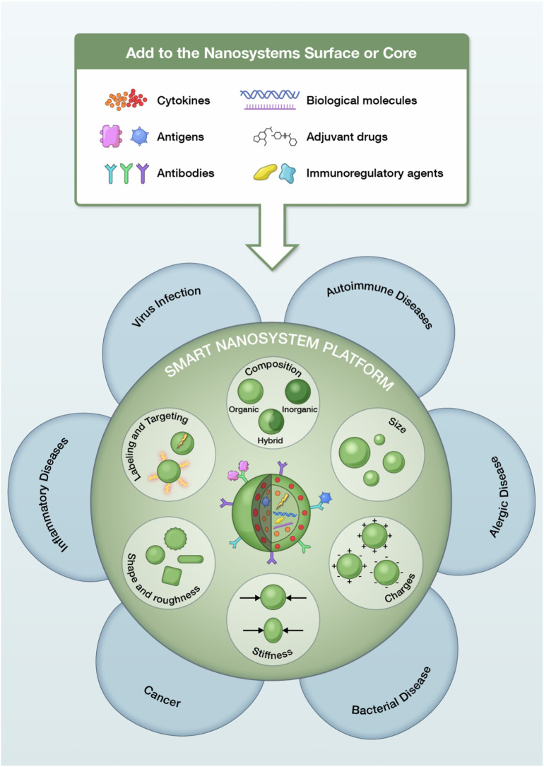
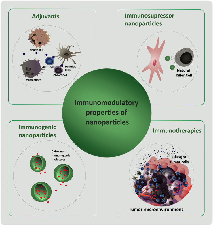
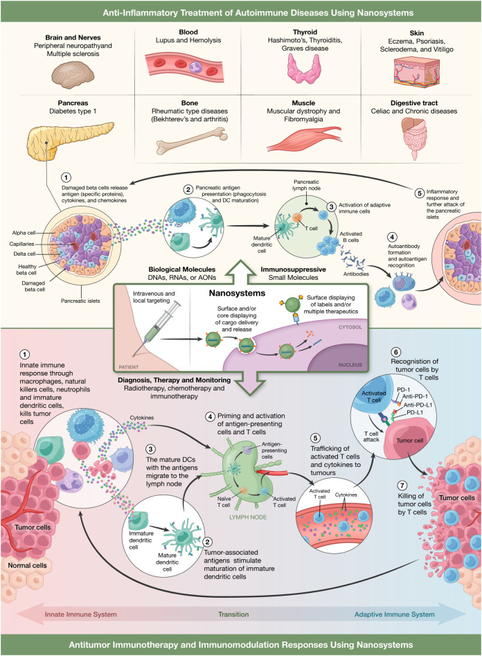
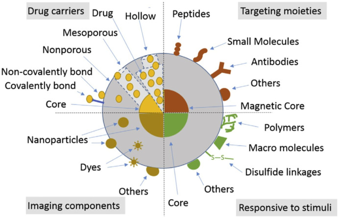
The development of nanoparticles (NPs) with potential therapeutic uses represents an area of vast interest in the scientific community during the last years. Recently, the pandemic caused by COVID-19 motivated a race for vaccines creation to overcome the crisis generated. This is a good demonstration that nanotechnology will most likely be the basis of future immunotherapy. Moreover, the number of publications based on nanosystems has significantly increased in recent years and it is expected that most of these developments can go on to experimentation in clinical stages soon. The therapeutic use of NPs to combat different diseases such as cancer, allergies or autoimmune diseases will depend on their characteristics, their targets, and the transported molecules. This review presents an in-depth analysis of recent advances that have been developed in order to obtain novel nanoparticulate based tools for the treatment of allergies, autoimmune diseases and for their use in vaccines. Moreover, it is highlighted that by providing targeted delivery an increase in the potential of vaccines to induce an immune response is expected in the future. Definitively, the here gathered analysis is a good demonstration that nanotechnology will be the basis of future immunotherapy.

Keywords: Allergy; Autoimmune disease; Immune stimulation; Immunomodulation therapy; Immunosuppressants; Nanoparticles.


Conflict of interest statement

The authors declare that they have no known competing financial interests or personal relationships that could have appeared to influence the work reported in this paper.

Figures

Fig. 1
Fig. 1
Schematic representation of different smart nanosystems and several biological molecules that can be used in the treatment of diverse pathologies.
Fig. 2
Fig. 2
Scheme of the NPs characteristics that can be exploited to modulate the immune system response.
Fig. 3
Fig. 3
Representation of anti-inflammatory nanosystems for the treatment of different autoimmune diseases with special focus, as pathophysiological model, in type 1 diabetes progression (upper part of the figure) or as immunotherapeutics against cancer where it can be seen the different stages of innate and adaptive immunity where nanosystems can intervene (lower part of the figure).
Fig. 4
Fig. 4
Schematic representation of SiNPs. Types of SiNPs for delivering biologically active agents and drugs. Targeting moieties on the surface of SiNPs or magnetic composites. SiNPs responding to stimuli (e.g. pH, glutathione, magnetic field, light and temperature). SiNPs for optical, magnetic resonance and other bioimaging applications. “Reprinted from Mebert et al. Copyright (2017), with permission from Elsevier”.
Fig. 5
Fig. 5
Schematic illustrations showing the composition/structure of the dual-responsive NPs developed in this study and their extracellular/intracellular anti-inflammatory mechanisms. “Reprinted from Pu et al. Copyright (2014), with permission from American Chemical Society”.
Fig. 6
Fig. 6
Intracellular localization of TA-Ag/AuNPs. The Manders' coefficients for co-localization of TA-Ag/AuNPs and cytoplasm (A) or lysosomes (B) in JAWS II cell culture exposed to 10 nm, 37 nm, 59 nm TA-AgNPs and 10 nm, 34 nm, 62 nm TA-AuNPs for 24 h at 2.5 μg/ml. *Significant differences with p ≤ 0.05. (C) Representative images for lysosomes (green), NPs (red) and nuclei (blue) in cells exposed to NPs, as described above. (D) NPs content in cells subjected to pretreatment with 10 μg/ml monodansylcadeverine and 5 μg/ml cytochalasin D, and then to incubation with TA-Ag/AuNPs at 2.5 μg/ml for 6 h. Reprinted from with permission from Frontiers under the terms of the Creative Commons Attribution License (CC BY).
Fig. 7
Fig. 7
(A–D) TEM micrographs of mature DC (mDC). (A) Untreated mDC. (B, C, D) mDC treated with 100 μg/ml of DC-SIGN pSiNP and cultured for 30 min, 2 h and 24 h. Arrows indicate surface binding and internalization of pSiNP. Scale barre presents 2 μm. (E–G) Fluorescence microscopy of mDC. (E) Untreated mDC. mDC cultured with 100 μg/ml of FITC-labelled isotype pSiNP (F) or DCSIGNpSiNP (G) taken at 24 h. Scale bar represents 40 μm at 40× magnification. (H) Flow cytometry histograms representing NPs uptake was dependent on DC-SIGN display. Monocyte-derived DCs treated with 20 μg/ml or 50 μg/ml of isotype pSiNP (black line) or DC-SIGN pSiNP (blue shaded)at 30 min, 2 h and 24 h. (n = 9, data is representative of one blood donor). Dashed line represents untreated DC control. Histograms show mean fluorescence intensity (MFI) and % positivity in parentheses. Reproduced from with permission from Elsevier.

References

    1. Medzhitov R., Janeway C.A. Innate immunity: impact on the adaptive immune response. Curr. Opin. Immunol. 1997;9:4–9. doi: 10.1016/S0952-7915(97)80152-5. - DOI - PubMed
    1. Nicholson L.B. The immune system. Essays Biochem. 2016;60:275–301. doi: 10.1042/EBC20160017. - DOI - PMC - PubMed
    1. Flajnik M.F., Du Pasquier L. Evolution of innate and adaptive immunity: can we draw a line? Trends Immunol. 2004;25:640–644. doi: 10.1016/j.it.2004.10.001. - DOI - PubMed
    1. Ren H., He Y., Liang J., Cheng Z., Zhang M., Zhu Y., Hong C., Qin J., Xu X., Wang J. Role of liposome size, surface charge, and PEGylation on rheumatoid arthritis targeting therapy. ACS Appl. Mater. Interfaces. 2019;11:20304–20315. doi: 10.1021/acsami.8b22693. - DOI - PubMed
    1. Cheng T., Zhang Y., Liu J., Ding Y., Ou H., Huang F., An Y., Liu Y., Liu J., Shi L. Ligand-switchable micellar nanocarriers for prolonging circulation time and enhancing targeting efficiency. ACS Appl. Mater. Interfaces. 2018;10:5296–5304. doi: 10.1021/acsami.7b18137. - DOI - PubMed

LinkOut - more resources