. 2022 Apr 29;8(17):eabj7892.
doi: 10.1126/sciadv.abj7892. Epub 2022 Apr 27.

A unified 3D map of microscopic architecture and MRI of the human brain

Affiliations

A unified 3D map of microscopic architecture and MRI of the human brain

Anneke Alkemade et al. Sci Adv. .

Abstract

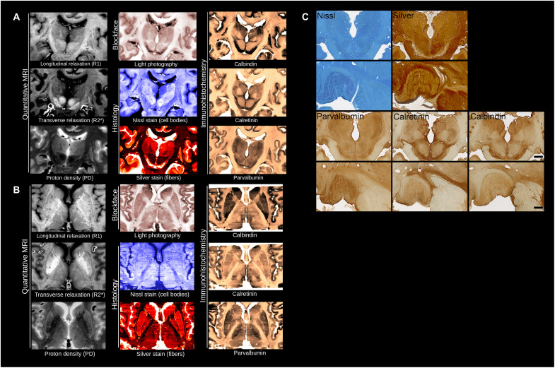
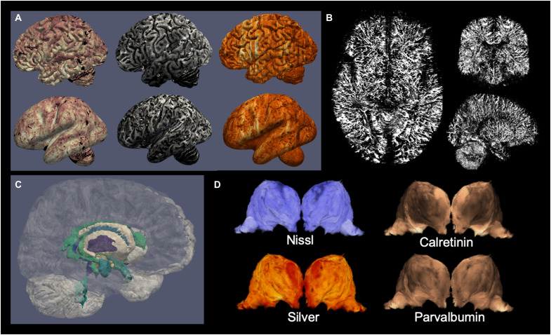
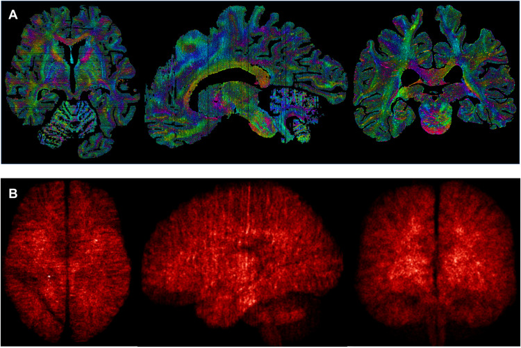
We present the first three-dimensional (3D) concordance maps of cyto- and fiber architecture of the human brain, combining histology, immunohistochemistry, and 7-T quantitative magnetic resonance imaging (MRI), in two individual specimens. These 3D maps each integrate data from approximately 800 microscopy sections per brain, showing neuronal and glial cell bodies, nerve fibers, and interneuronal populations, as well as ultrahigh-field quantitative MRI, all coaligned at the 200-μm scale to the stacked blockface images obtained during sectioning. These unprecedented 3D multimodal datasets are shared without any restrictions and provide a unique resource for the joint study of cell and fiber architecture of the brain, detailed anatomical atlasing, or modeling of the microscopic underpinnings of MRI contrasts.


Figures

Fig. 1.
Fig. 1.. Anatomical detail obtained from reconstructed human brains.
(A and B) Coronal (A) and axial view (B) for the three MRI contrasts, blockface images, and five microscopy stains. (C) Illustrations of single sections for Nissl and silver (Bielschowsky) staining, and parvalbumin, calretinin, and calbindin immunoreactivity in the thalamus (scale bar, 1 cm). High-power magnifications (bottom) show high-power magnifications of the lateral geniculate nucleus of the thalamus. Scale bar, 250 μm.
Fig. 2.
Fig. 2.. Examples of information derived from the dataset of specimen no. 15-2017.
(A) Cortical maps from the dataset: blockface (left), quantitative R2* (middle), and parvalbumin immunohistochemistry (right), sampled at the midcortical surface in fully folded (top) or inflated (bottom) views; (B) reconstructed blood vessels extracted from the coregistered stainings; (C) automated cortical and subcortical parcellations; (D) different stainings outline different thalamic nuclei boundaries.
Fig. 3.
Fig. 3.. Example of applications using the full range of available resolutions.
(A) Shape tensor analysis of the different sections at the 20-μm scale, recombined to the 200-μm space of the blockface image; (B) density maps of blood vessels detected from the background of the 20-μm sections realigned in 3D at 200 μm, projected, and averaged into MNI space at 0.5 mm.
Fig. 4.
Fig. 4.. Multistaining 2D-3D coregistration.
By acquiring consecutive blockface images, we reduce the registration problem to 2D-2D coregistration, which can provide good macroscopic alignment after parameter optimization (A). Coregistration across multiple stainings requires simultaneous alignment of each section to its neighbors and the blockface image (B). Using a forward-backward approach, we obtain smooth 3D reconstructions within three iterations of the registration procedure. Note that for each individual section, a separate blockface image was available, and neighboring stainings were only added to regularize the coregistration across slices and stainings. The registration pipeline code is publicly available (see Table 1).
Fig. 5.
Fig. 5.. Evaluation of microscopy slice registration in both subjects: Interslice distance between boundaries of the registered microscopy images, per marker and across markers.
Vertical dashed lines indicate the transitions between sampling strategies (see Table 1). Horizontal lines indicate the corresponding average interslice distance within markers. The >2-mm distances (high peaks) in the occipital pole (high slice numbers) reflect parts of the cerebellar cortex that were missing.

References

    1. Brodmann K., Neue Ergebnisse ueber die Vergleichende histologische Localisation der Grosshirnfinde mit besonderer Berucksichtigung des Stirnhirns. Anat. Anz. 41, 157–216 (1912).
    1. Vogt C., Vogt O., Die vergleichend-architektonische und die vergleichend-reizphysiologische Felderung der Großhirnrinde unter besonderer Berücksichtigung der menschlichen. Naturwissenchaften 14, 1190–1194 (1926).
    1. C. von Economo, G. N. Koskinas, Die Cytoarchitektonik der Hirnrinde des Erwachsenen Menschen (Springer, 1925).
    1. G. V. Childs, History of immunohistochemistry, in Pathobiology of Human Disease, L. M. McManus, R. N. Mitchell, Eds. (Elsevier, 2014).
    1. Nieuwenhuys R., Broere C. A. J., Cerliani L., A new myeloarchitectonic map of the human neocortex based on data from the Vogt–Vogt school. Brain Struct. Funct. 220, 2551–2573 (2015). - PubMed