. 2022 May 3;119(18):e2118126119.
doi: 10.1073/pnas.2118126119. Epub 2022 Apr 27.

Genomewide CRISPR knockout screen identified PLAC8 as an essential factor for SADS-CoVs infection

Affiliations

Genomewide CRISPR knockout screen identified PLAC8 as an essential factor for SADS-CoVs infection

Longping V Tse et al. Proc Natl Acad Sci U S A. .

Abstract

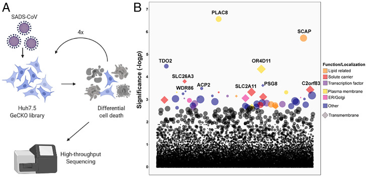
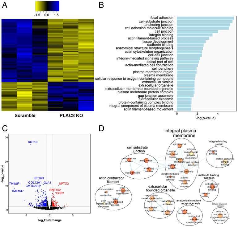
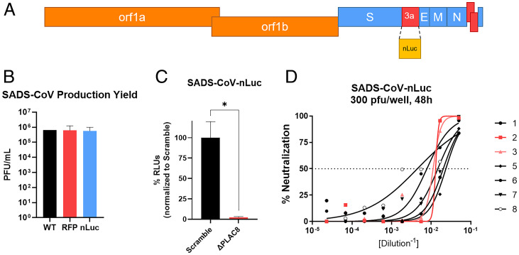
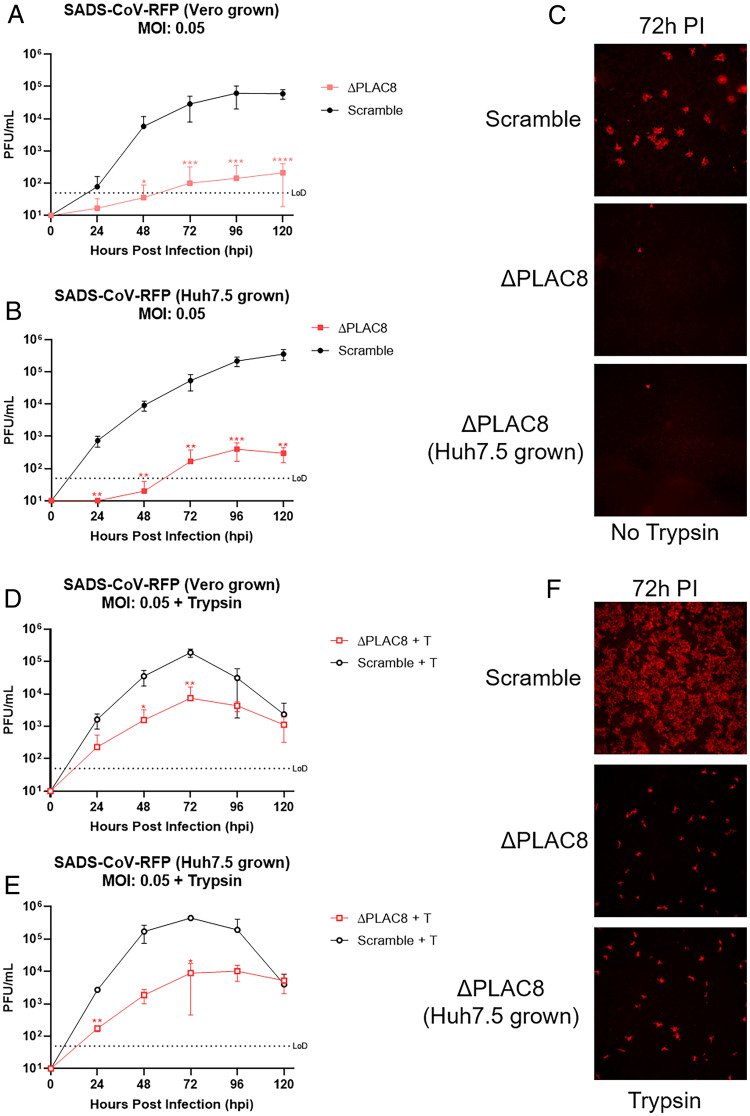
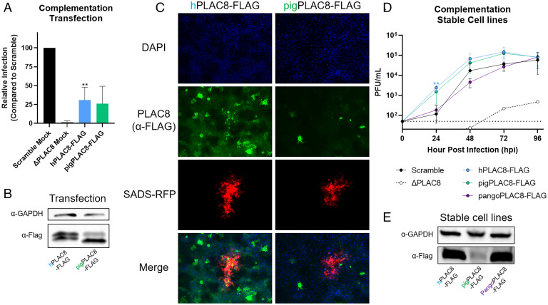
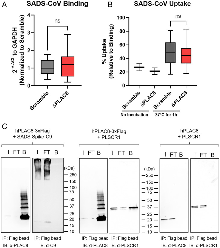
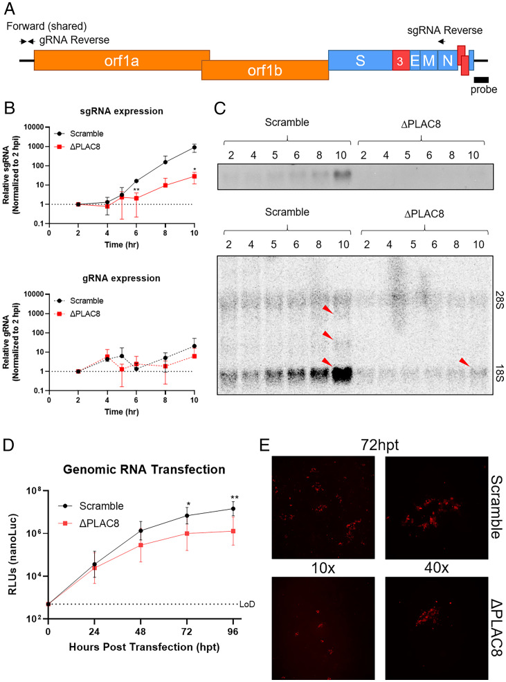
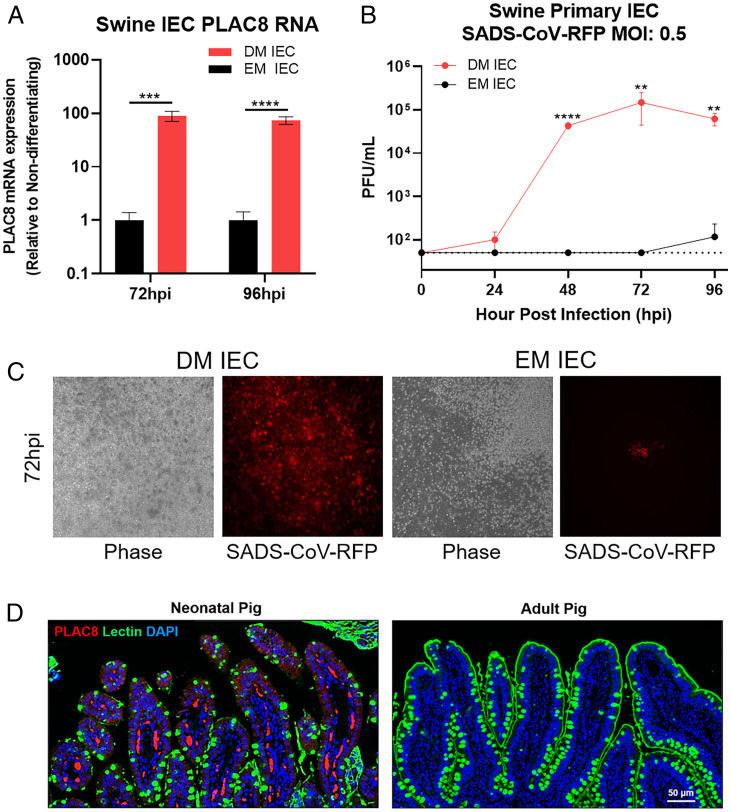
Zoonotic transmission of coronaviruses poses an ongoing threat to human populations. Endemic outbreaks of swine acute diarrhea syndrome coronavirus (SADS-CoV) have caused severe economic losses in the pig industry and have the potential to cause human outbreaks. Currently, there are no vaccines or specific antivirals against SADS-CoV, and our limited understanding of SADS-CoV host entry factors could hinder prompt responses to a potential human outbreak. Using a genomewide CRISPR knockout screen, we identified placenta-associated 8 protein (PLAC8) as an essential host factor for SADS-CoV infection. Knockout of PLAC8 abolished SADS-CoV infection, which was restored by complementing PLAC8 from multiple species, including human, rhesus macaques, mouse, pig, pangolin, and bat, suggesting a conserved infection pathway and susceptibility of SADS-CoV among mammals. Mechanistically, PLAC8 knockout does not affect viral entry; rather, knockout cells displayed a delay and reduction in viral subgenomic RNA expression. In a swine primary intestinal epithelial culture (IEC) infection model, differentiated cultures have high levels of PLAC8 expression and support SADS-CoV replication. In contrast, expanding IECs have low levels of PLAC8 expression and are resistant to SADS-CoV infection. PLAC8 expression patterns translate in vivo; the immunohistochemistry of swine ileal tissue revealed high levels of PLAC8 protein in neonatal compared to adult tissue, mirroring the known SADS-CoV pathogenesis in neonatal piglets. Overall, PLAC8 is an essential factor for SADS-CoV infection and may serve as a promising target for antiviral development for potential pandemic SADS-CoV.

Keywords: CRISPR; PLAC8; coronavirus; swine acute diarrhea syndrome coronavirus.


Conflict of interest statement

The authors declare no competing interest.

Figures

Fig. 1.
Fig. 1.
GeCKO library screening to identify critical genes involved in SADS-CoV infection. (A) Schematic of the CRISPR KO screening procedure on Huh7.5 cells. (B) Guide RNAs enriched after four rounds of SADS-CoV infection determined by high-throughput sequencing.
Fig. 2.
Fig. 2.
Transcriptomic analysis of PLAC8 KO Huh7.5 cells. (A) Heatmap of the normalized counts of significant DEGs between Huh7.5 Scramble and PLAC8 KO cells. Yellow indicates relatively lower expression, and blue indicates relatively higher expression. Data are shown from three replicate samples from each experimental condition. (B) Bar plot of the significantly enriched GO pathways between Huh7.5 Scramble and PLAC8 KO as determined by g:Profiler. (C) Volcano plot of DEGs between Scramble and PLAC8 KO Huh7.5 cells. Red denotes significantly up-regulated genes, and blue denotes significantly down-regulated genes. (D) Cytoscape EnrichmentMap of significantly enriched GO pathways as determined by g:Profiler.
Fig. 3.
Fig. 3.
Development of SADS-CoV-nLuc reporter virus via reverse genetics. (A) The design of SADS-CoV-nLuc virus with ORF3a replaced by nLuc. (B) Relative production yield of SADS-CoV infectious clone (WT), SADS-CoV-RFP (RFP), and SADS-CoV-nLuc (nLuc). (C) RLU/infectivity of SADS-CoV-nLuc (MOI: 0.0075) at 48 hpi in Scramble and PLAC8 KO Huh7.5 cells. (D) nLuc-based neutralization assay of SADS-CoV-nLuc against SADS-CoV Spike mouse antisera from eight mice. Error bars represent ± 1 SD, *P < 0.05 by Student's t test.
Fig. 4.
Fig. 4.
Growth kinetics of SADS-CoV-RFP. Infections were performed at a MOI of 0.05. (A) Growth kinetics of Vero-produced virus examined every 24 h for 120 hpi. (B) Growth kinetics of Huh7.5-produced virus examined every 24 h for 120 hpi. (C) Representative fluorescence images (4x) of SADS-CoV-RFP infection at 72 hpi. Growth kinetics of the virus were examined every 24 h for 120 hpi for virus produced in Vero cells (D) or Huh7.5 cells (E) in the presence of trypsin (1 µg/mL). (F) Representative fluorescence images (4x) of SADS-CoV-RFP infection in the presence of trypsin at 72 hpi. Error bars represent ± 1 SD, *P < 0.05, **P < 0.01, ***P < 0.001 by Student's t test.
Fig. 5.
Fig. 5.
Complementation with ectopic or stable PLAC8 expression rescues SADS-CoV infection. (A) PLAC8 KO cells were transiently transfected with human and pigPLAC8-FLAG (codon optimized). Subsequently, transfected cells were infected with SADS-CoV-nLuc (MOI: 0.015) for 48 hpi. RLUs from the infected cells were used as surrogates for viral infection. (B) Western blot images of hPLAC8 and pigPLAC8-FLAG expression on Huh7.5 cells after transient transfection using anti-FLAG antibody. (C) Representative immunofluorescence images of hPLAC8- and pigPLAC8-FLAG–transfected cells infected with SADS-CoV-RFP and stained with anti-FLAG antibody (green), SADS-CoV-RFP (red), and DAPI (blue). (D) SADS-CoV-RFP growth kinetics on Huh7.5 ΔPLAC8 cells stably expressing hPLAC8, codon-optimized pigPLAC8, and pangoPLAC8-FLAG. (E) Western blot images of hPLAC8, pigPLAC8, and pangoPLAC8-FLAG expression in stable cell lines using anti-FLAG antibody. Error bars represent ± 1 SD, *P < 0.05, **P < 0.01, ***P < 0.001 by Student's t test.
Fig. 6.
Fig. 6.
PLAC8 does not affect binding and internalization of SADS-CoV. Binding (A) and uptake (B) assay of SADS-CoV on Scramble and PLAC8 KO cells. For binding, cells were incubated with virus (MOI: 0.5) at 4 °C for 1 h and washed three times with HBSS. For uptake, after the wash from the binding assay, cells were incubated at 37 °C for 1 h. Surface virions were removed by 1) trypsin wash, 2) pH 2.5 acid wash, and 3) HBSS wash. Total RNA was isolated, and viral gRNA was determined by qRT-PCR. Data were plotted in a box-and-whisker plot, with the boxes representing 95% CI and whiskers representing maximum and minimum values from four independent experiments with three replicates. (C) Immunoprecipitation of PLAC8 with SADS-CoV spike protein. hPLAC8-FLAG and SADS-CoV spike with a C-terminal C9 tag were cotransfected in 293T cells for 48 h. Magnetic beads against FLAG tag were used for IP and blotted with α-PLAC8 or α-C9 antibodies. PLSCR1, a known factor that interacts with PLAC8, was used as the positive control, and PLAC8 without a FLAG tag was used as the negative control. I, input; FT, flow through; B, bound; ns, not significant; IP, immunoprecipitation; IB, immunoblot.
Fig. 7.
Fig. 7.
PLAC8 reduces viral sgRNA expression during infection and does not affect translation. (A) Schematic of SADS-CoV viral genome, qPCR primer sets, and Northern probe to determine gRNA and sgRNA expression. (B) Kinetics of gRNA and sgRNA expression of SADS-CoV-RFP from 2 to 10 hpi on Scramble and PLAC8 KO cells. (C) Northern blot image of viral RNA from B using radiolabeled probes against the SADS-CoV-RFP C terminus. Red arrows indicate sgRNA bands. (D) Kinetics of nLuc protein expression after viral gRNA transfection into Scramble and PLAC8 KO cells over 4 d. (E) Representative fluorescence images (10 and 40x) of the RFP signal detected in Scramble (Top) and PLAC8 KO (Bottom) cells at 72 hpt. Error bars represent ± 1 SD, *P < 0.05, **P < 0.01, ***P < 0.001 by Student's t test.
Fig. 8.
Fig. 8.
SADS-CoV infection in primary swine IECs. (A) PLAC8 mRNA expression of SADS-CoV-RFP–infected IECs in DM or EM at 72 and 96 hpi. (B) Viral growth of SADS-CoV-RFP on primary swine IEC in DM or EM. (C) Representative images of SADS-CoV-RFP–infected IECs in DM and EM at 72 hpi. (D) PLAC8 and glycocalyx integrity in neonatal and adult pig ileum. Neonatal and adult swine ileal sections of naïve pigs were stained with PLAC8 (red), UEA lectin (green), and DAPI (blue). Scale bar is 50 µm. Error bars represent ± 1 SD, *P < 0.05, **P < 0.01, ***P < 0.001, ****P < 0.0001 by Student's t test.

References

    1. Gallagher T., Perlman S., Broad reception for coronavirus. Nature 495, 176–177 (2013). - PMC - PubMed
    1. Drosten C., et al. , Identification of a novel coronavirus in patients with severe acute respiratory syndrome. N. Engl. J. Med. 348, 1967–1976 (2003). - PubMed
    1. Zaki A. M., van Boheemen S., Bestebroer T. M., Osterhaus A. D. M. E., Fouchier R. A. M., Isolation of a novel coronavirus from a man with pneumonia in Saudi Arabia. N. Engl. J. Med. 367, 1814–1820 (2012). - PubMed
    1. Wu F., et al. , A new coronavirus associated with human respiratory disease in China. Nature 579, 265–269 (2020). - PMC - PubMed
    1. Zhou P., et al. , A pneumonia outbreak associated with a new coronavirus of probable bat origin. Nature 579, 270–273 (2020). - PMC - PubMed

MeSH terms

Supplementary concepts