. 2022 May 3;119(18):e2201433119.
doi: 10.1073/pnas.2201433119. Epub 2022 Apr 27.

Dromedary camel nanobodies broadly neutralize SARS-CoV-2 variants

Affiliations

Dromedary camel nanobodies broadly neutralize SARS-CoV-2 variants

Jessica Hong et al. Proc Natl Acad Sci U S A. .

Abstract

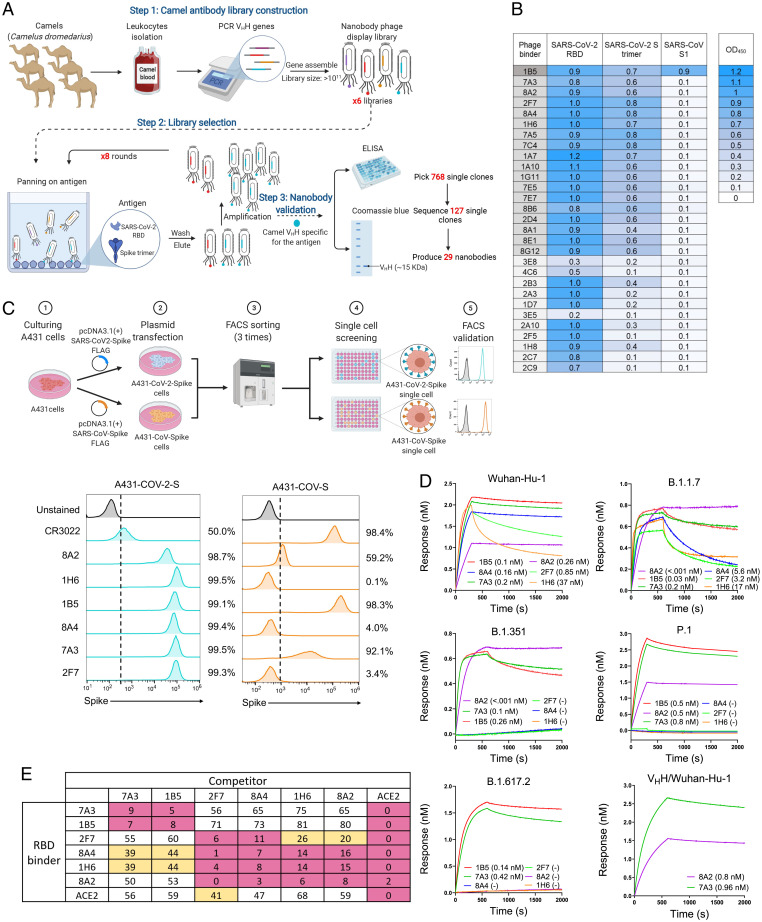
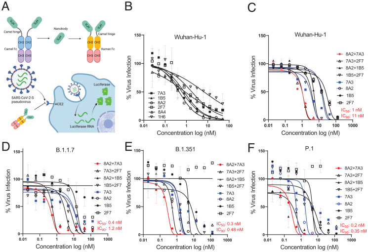
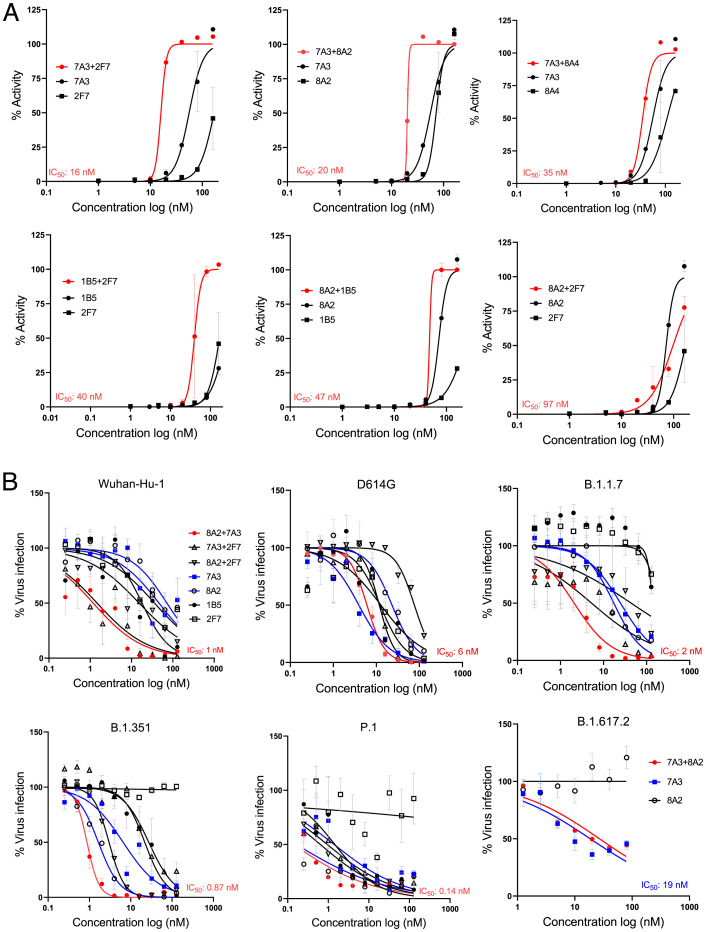
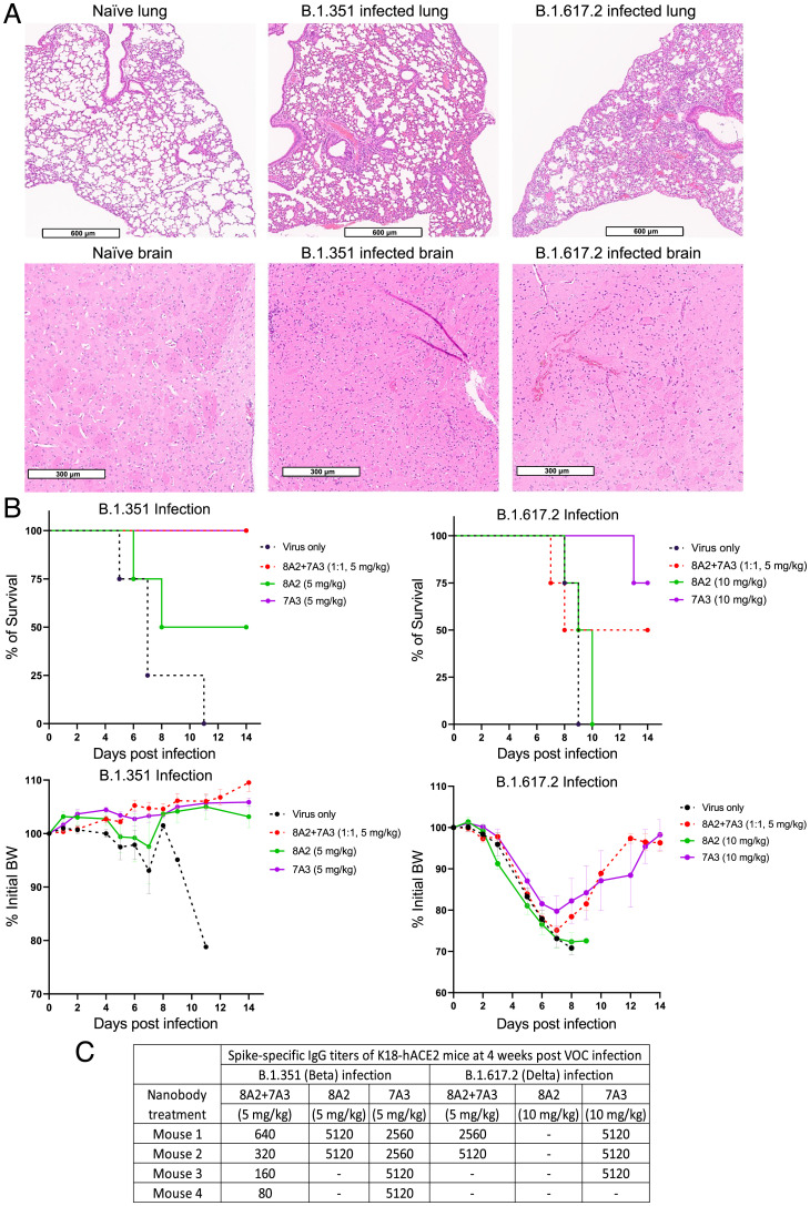
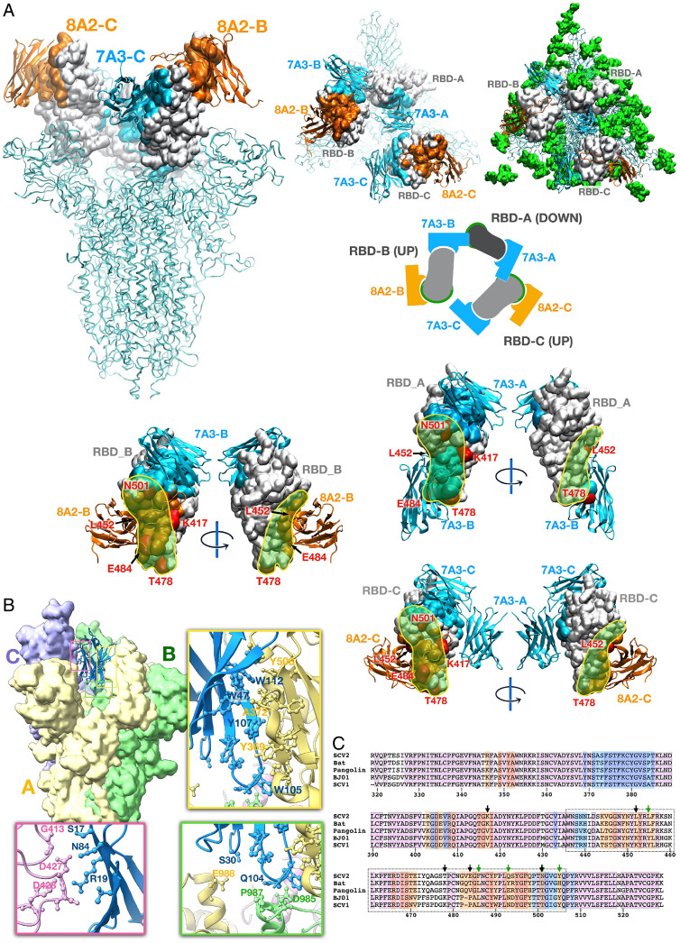
The severe acute respiratory syndrome coronavirus 2 (SARS-CoV-2) spike is a trimer of S1/S2 heterodimers with three receptor-binding domains (RBDs) at the S1 subunit for human angiotensin-converting enzyme 2 (hACE2). Due to their small size, nanobodies can recognize protein cavities that are not accessible to conventional antibodies. To isolate high-affinity nanobodies, large libraries with great diversity are highly desirable. Dromedary camels (Camelus dromedarius) are natural reservoirs of coronaviruses like Middle East respiratory syndrome CoV (MERS-CoV) that are transmitted to humans. Here, we built large dromedary camel VHH phage libraries to isolate nanobodies that broadly neutralize SARS-CoV-2 variants. We isolated two VHH nanobodies, NCI-CoV-7A3 (7A3) and NCI-CoV-8A2 (8A2), which have a high affinity for the RBD via targeting nonoverlapping epitopes and show broad neutralization activity against SARS-CoV-2 and its emerging variants of concern. Cryoelectron microscopy (cryo-EM) complex structures revealed that 8A2 binds the RBD in its up mode with a long CDR3 loop directly involved in the ACE2 binding residues and that 7A3 targets a deeply buried region that uniquely extends from the S1 subunit to the apex of the S2 subunit regardless of the conformational state of the RBD. At a dose of ≥5 mg/kg, 7A3 efficiently protected transgenic mice expressing hACE2 from the lethal challenge of variants B.1.351 or B.1.617.2, suggesting its therapeutic use against COVID-19 variants. The dromedary camel VHH phage libraries could be helpful as a unique platform ready for quickly isolating potent nanobodies against future emerging viruses.

Keywords: SARS-CoV-2; cryo-EM; dromedary camel nanobody VHH; neutralizing antibody; single-domain antibody.


Conflict of interest statement

Competing interest statement: M.H. and J.H. are inventors on provisional patent application no. US 63/105,769 assigned to the NIH, “Single domain antibodies targeting SARS coronavirus spike protein and uses thereof.”

Figures

Fig. 1.
Fig. 1.
Isolation of high-affinity camel nanobodies against SARS-CoV-2. (A) Isolation of camel VHHs that bind the RBD by phage display. (B) Camel VHHs against the S protein of SARS-CoV-2 or SARS-CoV. (C) Flow cytometry (FACS) was performed to monitor the cross-reaction of nanobodies to the spike of both SARS-CoV-2 and SARS-CoV on human cells. The cartoon outlines the experimental workflow for overexpression of SARS-CoV-2-spike or SARS-CoV-spike in the A431 human cell line. Both cell lines were stained with VHH nanobodies or CR3022 as a positive control. (D) Binding (KD) of VHH-hFc or VHHs against the RBD of SARS-CoV-2 and its variants. (E) Cross-competition assay of each nanobody and ACE2 on Octet.
Fig. 2.
Fig. 2.
Nanobodies neutralize SARS-CoV-2 and the variants in pseudovirus assay. (A) Diagrams illustrating pseudovirus assay and VHH-hFc. (B) Camel VHH-hFc proteins inhibit SARS-CoV-2 pseudovirus infectivity to ACE2 expressing human cells by measuring luciferase expression. (CF) Pseudovirus particle neutralization assay testing 2-in-1 combinations and single nanobodies showing that 7A3 + 8A2 combination has the best neutralization activity.
Fig. 3.
Fig. 3.
Neutralization of SARS-CoV-2 and its variants in live virus assay. (A) Live virus neutralization assay of nanobodies 7A3, 8A2, 2F7, 1B5, and 8A4 along with their 2-in-1 combinations against Wuhan-Hu-1. (B) Live variant virus assay using the top three nanobody combinations of 8A2 + 7A3, 8A2 + 2F7, and 7A3 + 2F7 was conducted against Wuhan-Hu-1, D614G, B.1.1.7, B.1.351, P.1, and B.1.617.2.
Fig. 4.
Fig. 4.
Protection of the K18-hACE2 mice infected with a lethal dose of B.1.351 or B.1.617.2 variant. (A) The histology of lung and brain tissues harvested from K18-hACE2 transgenic mice on day 6 or 7 following infection of live SARS-CoV-2 B.1.351 or B.1.617.2 variant virus. (B) K18-hACE2 mice (n = 4 per group) were injected intraperitoneally with nanobody 7A3, 8A2, or 2-in-1 mixture at indicated doses followed by a lethal infection of B.1.351 or B.1.617.2 strain. Mortality and BW were monitored for 2 wk postinfection. (C) Spike-specific IgG titers of K18-hACE2 mice surviving the lethal B.1.351 or B.1.617.2 infection. Dash denotes that mice died during infection.
Fig. 5.
Fig. 5.
Cryo-EM structure of 7A3 and 8A2 nanobodies with SARS-Cov-2 spike. (A) The structure models based on the EM images show that 8A2 and 7A3 bind two distinct sites on the RBD. The mutations on the RBD derived from B.1.1.7 (N501Y), B.1.351 (K417N, E484K, N501Y), P.1 (K417T, E484K, N501Y), and B.1.617.2 (K417N, L452R, and T478K) are indicated. Green spheres (Top Right) are glycans. (B) The unique 7A3 binding pattern. (C) Nanobodies epitopes coverage as determined from the experimental structure using a 5-Å contact cutoff. Sequences of coronavirus RBD for Bat_RaTG13 (A0A6B9WHD3), Human BJ01 (Q6GYR1), Pangolin (A0A6G6A2Q2), SARS-CoV-2 (P0DTC2), and SARS-CoV-1 (P59594) are presented for comparison purposes. Degree of sequence identity is indicated by background pink hue. The 7A3 contact is indicated in blue, and 8A2 is indicated in orange. Arrows indicate mutation sites of concern (black) and key ACE2 contact residues (green). RBM is indicated by the dotted box. PDB entry codes for our complex structures of the 7A3 and 8A2 VHHs with the SARS-CoV-2 stabilized spike: PDB ID 7TPR, EMD-26062.

Update of

References

    1. Li Q., et al. , Early transmission dynamics in Wuhan, China, of novel coronavirus-infected pneumonia. N. Engl. J. Med. 382, 1199–1207 (2020). - PMC - PubMed
    1. Zhou F., et al. , Clinical course and risk factors for mortality of adult inpatients with COVID-19 in Wuhan, China: A retrospective cohort study. Lancet 395, 1054–1062 (2020). - PMC - PubMed
    1. Vaduganathan M., et al. , Renin-angiotensin-aldosterone system inhibitors in patients with Covid-19. N. Engl. J. Med. 382, 1653–1659 (2020). - PMC - PubMed
    1. Yan R., et al. , Structural basis for the recognition of SARS-CoV-2 by full-length human ACE2. Science 367, 1444–1448 (2020). - PMC - PubMed
    1. Yuan M., et al. , A highly conserved cryptic epitope in the receptor binding domains of SARS-CoV-2 and SARS-CoV. Science 368, 630–633 (2020). - PMC - PubMed

Publication types

Substances

Supplementary concepts