. 2022 Apr 27;17(4):e0267155.
doi: 10.1371/journal.pone.0267155. eCollection 2022.

Dimerization of the pulmonary surfactant protein C in a membrane environment

Affiliations

Dimerization of the pulmonary surfactant protein C in a membrane environment

Hanna Korolainen et al. PLoS One. .

Abstract

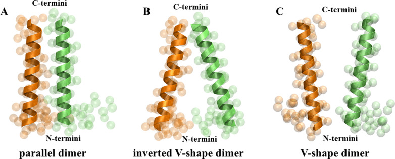
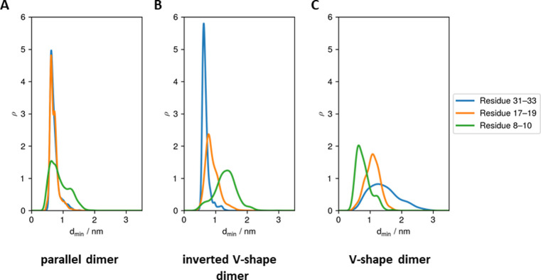
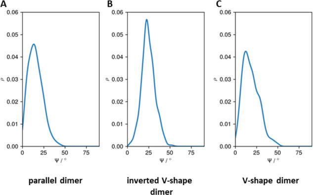
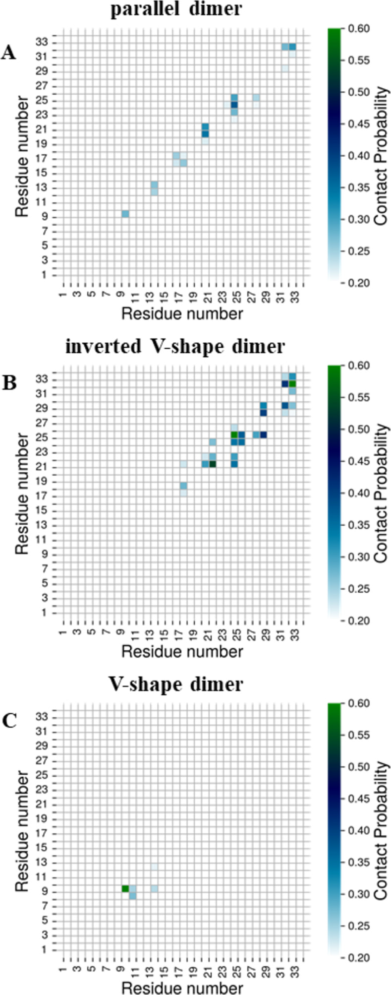
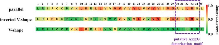
Surfactant protein C (SP-C) has several functions in pulmonary surfactant. These include the transfer of lipids between different membrane structures, a role in surfactant recycling and homeostasis, and involvement in modulation of the innate defense system. Despite these important functions, the structures of functional SP-C complexes have remained unclear. SP-C is known to exist as a primarily α-helical structure with an apparently unstructured N-terminal region, yet there is recent evidence that the functions of SP-C could be associated with the formation of SP-C dimers and higher oligomers. In this work, we used molecular dynamics simulations, two-dimensional umbrella sampling, and well-tempered metadynamics to study the details of SP-C dimerization. The results suggest that SP-C dimerizes in pulmonary surfactant membranes, forming dimers of different topologies. The simulations identified a dimerization motif region V21xxxVxxxGxxxM33 that is much larger than the putative A30xxxG34 motif that is commonly assumed to control the dimerization of some α-helical transmembrane domains. The results provide a stronger basis for elucidating how SP-C functions in concert with other surfactant proteins.


Conflict of interest statement

The authors have declared that no competing interests exist.

Figures

Fig 1
Fig 1
A) Atomistic (left) and coarse-grained (right) model of surfactant protein C (SP-C). B) Chemical structure of lipids used to mimic the pulmonary surfactant membrane: DPPC, POPC, POPG, and cholesterol (CHOL). See the text for details.
Fig 2
Fig 2. The definition of ϕ1 and ϕ2 dihedral angles.
The centers of mass (COMs) of the upper (A/A’), middle (B/B’), and lower (C/C’) groups of residues were picked as the three points forming each dihedral, while the fourth point (C’/C) belonged to the other monomer. The upper points (A/A’) were defined as the COM of LEU31, LEU32, and MET33 in each SP-C monomer. The middle points (B/B’) were defined as COM of VAL17, VAL18, and VAL19. Finally, the lowest points (C/C’) were defined as COM of VAL8, ASN9, and LEU10.
Fig 3
Fig 3
A) The SP-C dimerization free energy as a function of the minimum distance between any two beads belonging to SP-C monomers. Structures with dmin smaller or equal to 0.60 nm (depicted as black dashed line) are considered the bound dimers. We marked the untapped region of the free energy profile with a blue dashed line. B) The average minimum distance between N-termini (residues 8–10; green line), middles of helices (residues 17–19; orange line), and C-termini (residues 31–33; blue line) of SP-C monomers as a function of the COM-COM distance between SP-C monomers.
Fig 4
Fig 4
Representative structures (centroids) of three distinctive topologies of the SP-C homodimer ordered by the increasing free energy values: (A) parallel dimer, (B) inverted V-shape dimer, and (C) V-shape dimer. The SP-C monomers involved in the formation of homodimers are colored differently (orange and lime) for clarity.
Fig 5
Fig 5
The distributions of the minimum distances between N-termini (green line), middles of helices (orange line), and C-termini (blue line) of SP-C monomers in each dimer topology: A) parallel dimer, B) inverted V-shape dimer, and C) V-shape dimer.
Fig 6
Fig 6
The distributions of the crossing angles (Ψ) between the helices forming the homodimer in each dimer topology: A) parallel dimer, B) inverted V-shape dimer, and C) V-shape dimer.
Fig 7
Fig 7. Contact probability maps for each pair of residues in the SP-C homodimers averaged over all homodimers belonging to the given dimer topology.
Contact probabilities equal to one correspond to the situation where given residues are in contact in all homodimers belonging to this cluster topology. Two residues were considered to be in contact, if any of their beads were closer than 6 Å. Only contact probabilities higher than 0.20 are shown for clarity.
Fig 8
Fig 8. Contact probabilities of SP-C residues for all dimer topologies identified in this study.
The purple dashed line depicts the position of the putative AxxxG motif as reported by Kairys et al. [23].

References

    1. Singh SK. Human respiratory viral infections: CRC Press; 2014.
    1. Goerke J. Pulmonary surfactant: Functions and molecular composition. Biochim Biophys Acta. 1998;1408(2–3):79–89. doi: 10.1016/s0925-4439(98)00060-x - DOI - PubMed
    1. Lopez-Rodriguez E, Pérez-Gil J. Structure-function relationships in pulmonary surfactant membranes: From biophysics to therapy. Biochim Biophys Acta. 2014;1838(6):1568–85. doi: 10.1016/j.bbamem.2014.01.028 - DOI - PubMed
    1. Pérez-Gil J. Structure of pulmonary surfactant membranes and films: The role of proteins and lipid–protein interactions. Biochim Biophys Acta. 2008;1778(7–8):1676–95. doi: 10.1016/j.bbamem.2008.05.003 - DOI - PubMed
    1. Perez-Gil J, Casals C, Marsh D. Interactions of hydrophobic lung surfactant proteins SP-B and SP-C with dipalmitoylphosphatidylcholine and dipalmitoylphosphatidylglycerol bilayers studied by electron spin resonance spectroscopy. Biochemistry. 1995;34(12):3964–71. doi: 10.1021/bi00012a014 - DOI - PubMed

Publication types

MeSH terms

Substances