

SARS-CoV-2 variants C.1.2 and B.1.621 (Mu) partially evade neutralization by antibodies elicited upon infection or vaccination
- PMID: 35477025
- PMCID: PMC9010234
- DOI: 10.1016/j.celrep.2022.110754
SARS-CoV-2 variants C.1.2 and B.1.621 (Mu) partially evade neutralization by antibodies elicited upon infection or vaccination
Abstract
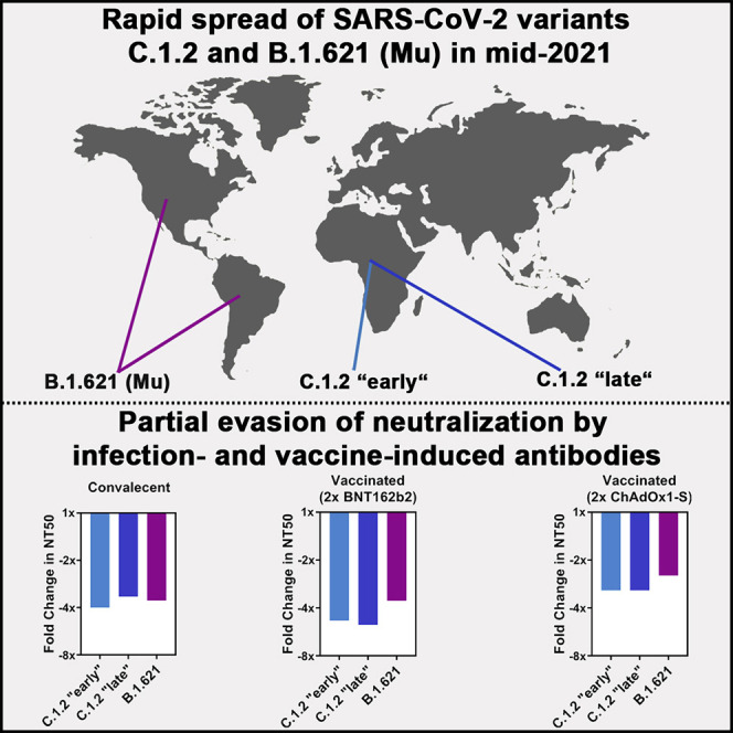
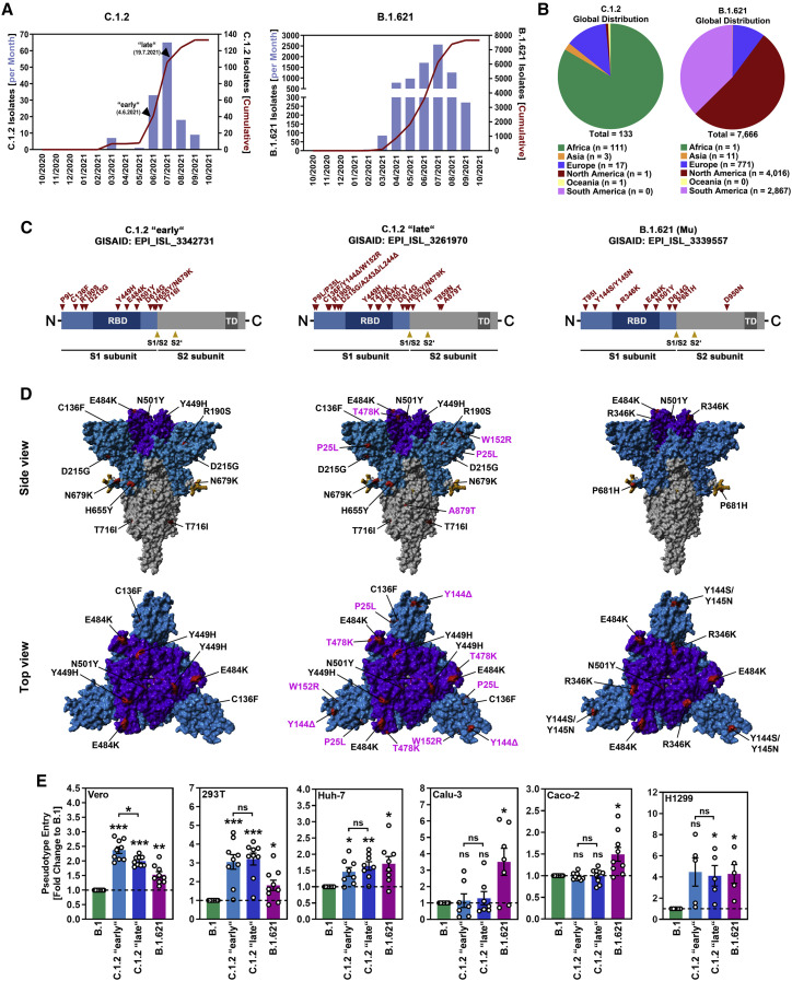
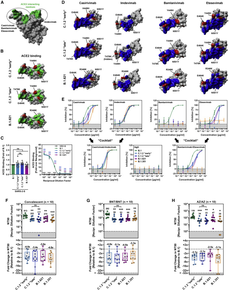
Rapid spread of SARS-CoV-2 variants C.1.2 and B.1.621 (Mu variant) in Africa and the Americas, respectively, as well as a high number of mutations in the viral spike proteins raised concerns that these variants might pose an elevated threat to human health. Here, we show that C.1.2 and B.1.621 spike proteins mediate increased entry into certain cell lines but do not exhibit increased ACE2 binding. Further, we demonstrate that C.1.2 and B.1.621 are resistant to neutralization by bamlanivimab but remain sensitive to inhibition by antibody cocktails used for COVID-19 therapy. Finally, we show that C.1.2 and B.1.621 partially escape neutralization by antibodies induced upon infection and vaccination, with escape of vaccine-induced antibodies being as potent as that measured for B.1.351 (Beta variant), which is known to be highly neutralization resistant. Collectively, C.1.2 and B.1.621 partially evade control by vaccine-induced antibodies, suggesting that close monitoring of these variants is warranted.
Keywords: B.1.621; C.1.2; COVID-19; CP: Immunology; CP: Microbiology; Mu; SARS-CoV-2; antibody; neutralization; spike; variants.
Copyright © 2022 The Author(s). Published by Elsevier Inc. All rights reserved.
Conflict of interest statement
Declaration of interests M.S.W. received unrestricted funding for independent research projects from Sartorius.
Figures
Similar articles
-
Decreased and Heterogeneous Neutralizing Antibody Responses Against RBD of SARS-CoV-2 Variants After mRNA Vaccination.Front Immunol. 2022 Apr 6;13:816389. doi: 10.3389/fimmu.2022.816389. eCollection 2022. Front Immunol. 2022. PMID: 35464418 Free PMC article.
-
Comprehensive characterization of the antibody responses to SARS-CoV-2 Spike protein finds additional vaccine-induced epitopes beyond those for mild infection.Elife. 2022 Jan 24;11:e73490. doi: 10.7554/eLife.73490. Elife. 2022. PMID: 35072628 Free PMC article.
-
Serum Neutralizing Activity of mRNA-1273 against SARS-CoV-2 Variants.J Virol. 2021 Nov 9;95(23):e0131321. doi: 10.1128/JVI.01313-21. Epub 2021 Sep 22. J Virol. 2021. PMID: 34549975 Free PMC article. Clinical Trial.
-
Variants of SARS-CoV-2, their effects on infection, transmission and neutralization by vaccine-induced antibodies.Eur Rev Med Pharmacol Sci. 2021 Sep;25(18):5857-5864. doi: 10.26355/eurrev_202109_26805. Eur Rev Med Pharmacol Sci. 2021. PMID: 34604978 Review.
-
Antibody-mediated neutralization of SARS-CoV-2.Immunity. 2022 Jun 14;55(6):925-944. doi: 10.1016/j.immuni.2022.05.005. Epub 2022 May 13. Immunity. 2022. PMID: 35623355 Free PMC article. Review.
Cited by
-
A SARS-CoV-2: Companion Animal Transmission and Variants Classification.Pathogens. 2023 May 29;12(6):775. doi: 10.3390/pathogens12060775. Pathogens. 2023. PMID: 37375465 Free PMC article. Review.
-
Challenges in Emerging Vaccines and Future Promising Candidates against SARS-CoV-2 Variants.J Immunol Res. 2024 Jan 25;2024:9125398. doi: 10.1155/2024/9125398. eCollection 2024. J Immunol Res. 2024. PMID: 38304142 Free PMC article. Review.
-
Nanomaterials to combat SARS-CoV-2: Strategies to prevent, diagnose and treat COVID-19.Front Bioeng Biotechnol. 2022 Nov 25;10:1052436. doi: 10.3389/fbioe.2022.1052436. eCollection 2022. Front Bioeng Biotechnol. 2022. PMID: 36507266 Free PMC article. Review.
-
Cross-reactive humoral and CD4+ T cell responses to Mu and Gamma SARS-CoV-2 variants in a Colombian population.Front Immunol. 2023 Jul 27;14:1241038. doi: 10.3389/fimmu.2023.1241038. eCollection 2023. Front Immunol. 2023. PMID: 37575243 Free PMC article.
-
The SARS-CoV-2 Delta-Omicron Recombinant Lineage (XD) Exhibits Immune-Escape Properties Similar to the Omicron (BA.1) Variant.Int J Mol Sci. 2022 Nov 14;23(22):14057. doi: 10.3390/ijms232214057. Int J Mol Sci. 2022. PMID: 36430535 Free PMC article.
References
MeSH terms
Substances
Supplementary concepts
LinkOut - more resources
Full Text Sources
Other Literature Sources
Medical
Research Materials
Miscellaneous
