Case Reports
. 2022 Jun 28;6(12):3779-3791.
doi: 10.1182/bloodadvances.2022007029.

Functional interaction between compound heterozygous TERT mutations causes severe telomere biology disorder

Affiliations
Case Reports

Functional interaction between compound heterozygous TERT mutations causes severe telomere biology disorder

Aram Niaz et al. Blood Adv. .

Abstract

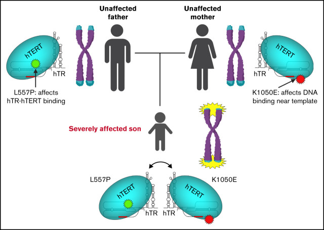
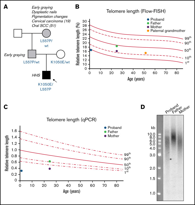
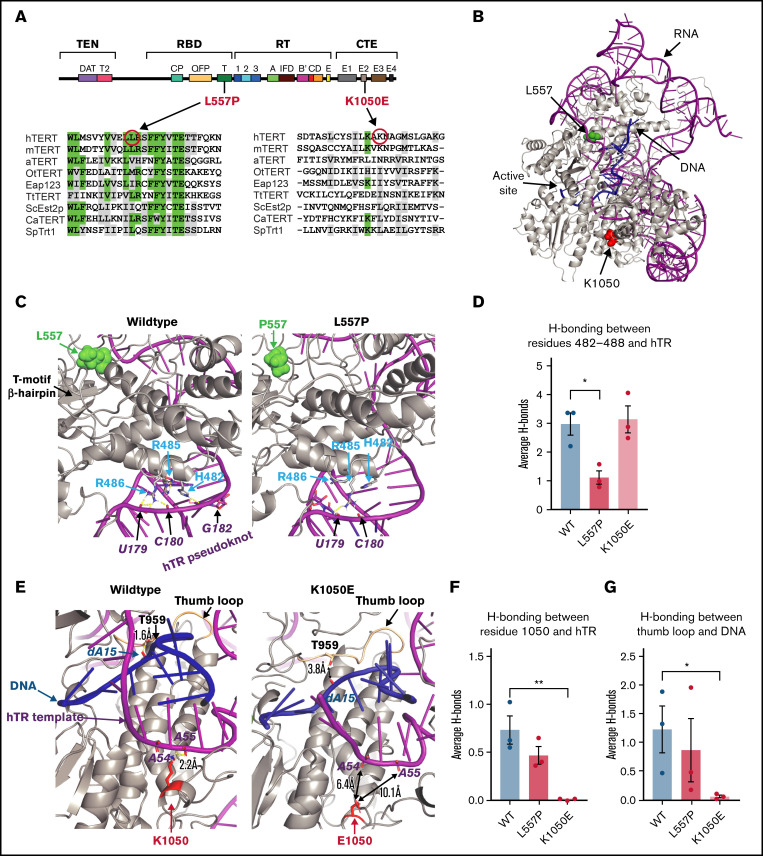
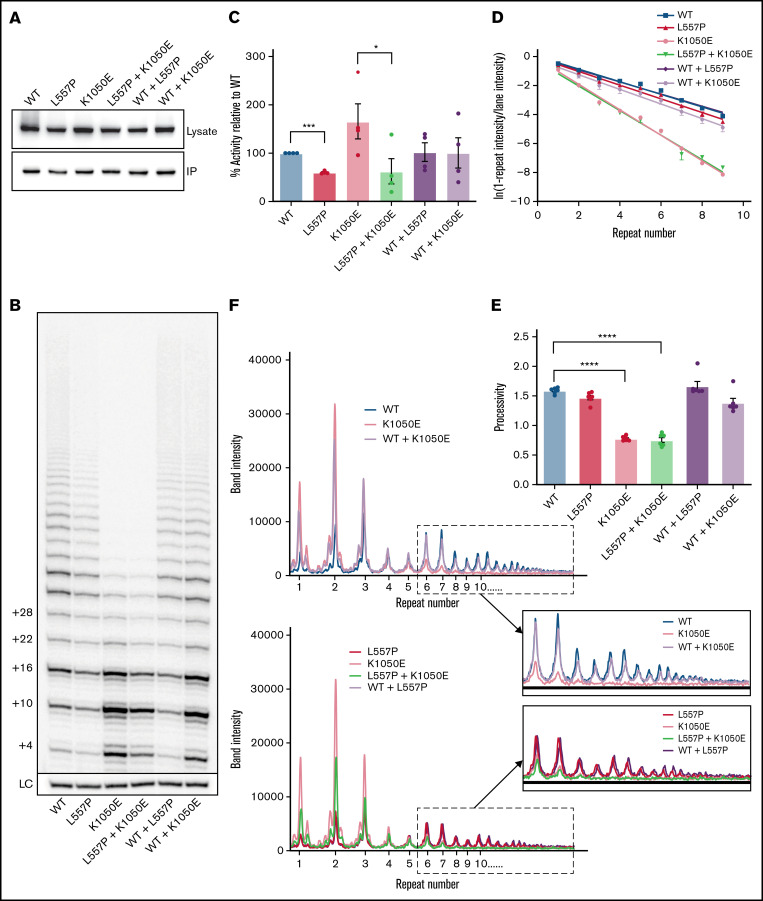
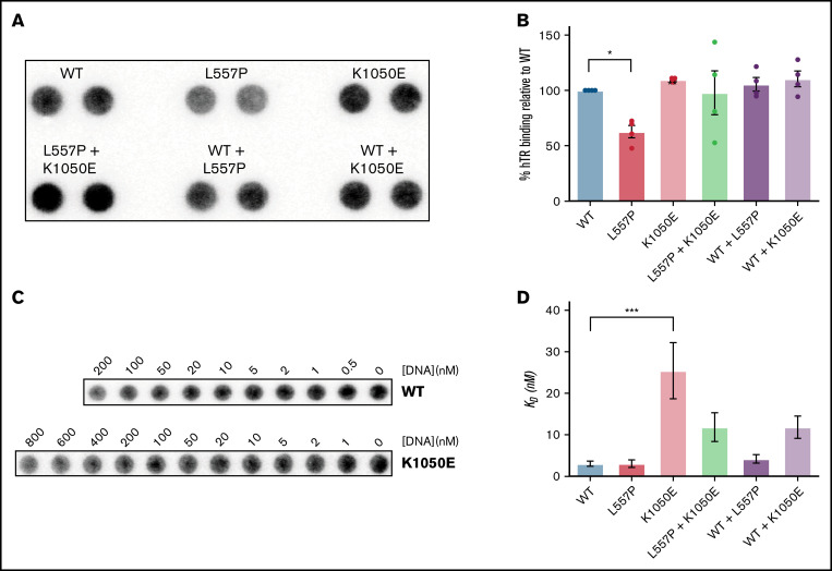
Telomere biology disorders (TBDs) are a spectrum of multisystem inherited disorders characterized by bone marrow failure, resulting from mutations in the genes encoding telomerase or other proteins involved in maintaining telomere length and integrity. Pathogenicity of variants in these genes can be hard to evaluate, because TBD mutations show highly variable penetrance and genetic anticipation related to inheritance of shorter telomeres with each generation. Thus, detailed functional analysis of newly identified variants is often essential. Herein, we describe a patient with compound heterozygous variants in the TERT gene, which encodes the catalytic subunit of telomerase, hTERT. This patient had the extremely severe Hoyeraal-Hreidarsson form of TBD, although his heterozygous parents were clinically unaffected. Molecular dynamic modeling and detailed biochemical analyses demonstrate that one allele (L557P) affects association of hTERT with its cognate RNA component hTR, whereas the other (K1050E) affects the binding of telomerase to its DNA substrate and enzyme processivity. Unexpectedly, the data demonstrate a functional interaction between the proteins encoded by the two alleles, with wild-type hTERT rescuing the effect of K1050E on processivity, whereas L557P hTERT does not. These data contribute to the mechanistic understanding of telomerase, indicating that RNA binding in one hTERT molecule affects the processivity of telomere addition by the other molecule. This work emphasizes the importance of functional characterization of TERT variants to reach a definitive molecular diagnosis for patients with TBD, and, in particular, it illustrates the importance of analyzing the effects of compound heterozygous variants in combination, to reveal interallelic effects.


Figures

None
Graphical abstract
Figure 1.
Figure 1.
Family pedigree and telomere length measurements of patient with Hoyeraal-Hreidarsson syndrome. (A) Pedigree of family in which the proband has compound heterozygous variants in TERT. Amino acid changes resulting from the TERT variants are labeled in blue. Arrow and black shaded box indicate the proband with Hoyeraal-Hreidarsson syndrome; gray shading indicates those with mild DC features. (B) Relative telomere length (as a percentage of that of the control 4n cell line CCRF-CEM) according to age, measured by Flow-FISH. Lines represent the 1st, 10th, 50th, 90th, and 99th percentiles of telomere length of 240 healthy controls. (C) Relative telomere length according to age, measured by qPCR. Lines represent the 1st, 10th, 50th, 90th, and 99th percentiles of telomere length of 240 healthy controls. (D) Telomere length in the proband and his parents, measured by Southern blot analysis with a probe against telomeric DNA.
Figure 2.
Figure 2.
Molecular modeling predicts that L557P affects hTERT binding to hTR, whereas K1050E affects DNA binding. (A) Schematic of the secondary structure of hTERT, showing the 4 major functional domains: telomerase essential N-terminal (TEN) domain, RNA binding domain (RBD), reverse transcriptase (RT) domain, and C-terminal extension (CTE) domain. The alignments show the degree of conservation of the regions surrounding L557P and K1050E in 9 species, including vertebrates, a plant, ciliated protozoa, and yeast: hTERT, human; mTERT, Mus musculus; aTERT, Arabidopsis thaliana; OtTERT, Oxytricha trifallax; Eap123, Euplotes aediculatus; TtTERT, Tetrahymena thermophila; ScEst2p, Saccharomyces cerevisiae; CaTERT, Candida albicans; SpTRT1p, Schizosaccharomyces pombe. Identical positions are indicated in green; positions showing amino acid similarity are highlighted in gray. (B) Model of WT hTERT used for molecular dynamics simulations. Protein, RNA, and DNA are gray, magenta, and blue, respectively. Locations of investigated mutations, L557 and K1050, are represented as space-filling atoms and colored in green and red, respectively. (C) Effect of L557P (green spheres): close-up view of the shift in position of the 482-to-488 helix relative to hTR in hTERTL557P (right) compared with WT hTERT (left). The change in hydrogen bonding between the 482-to-488 loop and hTR is quantified in panel D; data are the mean ± standard error of the mean (SEM; n = 3). *P = .0147, by 2-tailed t test. (E) Effect of K1050E: in hTERTwt (gray, left), K1050 (red) forms a hydrogen bond with hTR (magenta) at adenine-55. This hydrogen bond is lost in the hTERTK1050E protein (right) (quantified in panel F; data are the mean ± SEM [n = 3]; **P = .0074, by 2-tailed t test). In the hTERTK1050E simulation, the DNA substrate (residues 11-18) is shifted away from the thumb loop (hTERT residues 957-965; wheat) compared with WT and is accompanied by a reduction in hydrogen bonding between the thumb loop and DNA in hTERTK1050E compared with WT (quantified in panel G; data are the mean ± SEM [n = 3]; *P = .0461, by 2-tailed t test). Hydrogen bonds are yellow dashed lines (E), and the distance between a protein residue and the nearest nucleotides (outside hydrogen bonding distances) are indicated by double-headed black arrows (E; right).
Figure 3.
Figure 3.
The effects of L557P and K1050E on telomerase activity. (A) Western blot analyses of HEK293T cell lysates expressing the indicated hTERT variants (top) and the corresponding samples purified by IP (bottom). Blots were probed with an anti-FLAG antibody to detect exogenous FLAG-tagged hTERT. (B) Direct activity assay showing extension of a telomeric DNA primer in vitro in the presence of radiolabeled 32P-dGTP, for WT telomerase and the indicated variants, expressed individually or together. The number of nucleotides added to the primer is shown on the left. LC: 32P-labeled 30-mer oligonucleotide included as a control for recovery and loading. (C) Quantitation of total telomerase activity relative to WT for the indicated variants or combinations. Data are the mean ± standard error of the mean (SEM; n = 4). *P = .042; ***P = .0003, by repeated-measures 1-way analysis of variance (ANOVA), followed by Dunnett’s post hoc test. (D) Calculation of the amount of telomerase processivity for the indicated hTERT variants and combinations. For each telomere repeat added by telomerase, the logs of the “fraction of products left behind” (ie, dissociated from the enzyme) were plotted against the repeat number, and the plot was fitted by linear regression, with the slope inversely proportional to processivity. Each data point represents the mean ± SEM (n = 6). (E) Processivity values (defined as −0.693/m, where m is the slope of the line) were calculated from the 6 individual experiments shown in panel D. Data are the mean ± SEM (n = 6). ****P < .0001, by repeated measures 1-way ANOVA, followed by Dunnett’s post hoc test. (F) Line graphs of the intensities of bands in the indicated lanes of the gel in panel B. WT and K1050E telomerase, expressed individually or together (top). L557P and K1050E telomerase, expressed individually or together (bottom). Magnification of the boxed regions of the plots, showing repeat 6 and higher (inset).
Figure 4.
Figure 4.
L557P causes a defect in telomerase assembly, whereas K1050E affects binding of telomerase to its DNA substrate. (A) Northern dot-blot showing the amount of hTR recovered after IP of hTERT from lysates of HEK293T cells expressing the indicated hTERT variants. Samples were normalized to the recovery of hTERT on western blots and loaded in duplicate, with their average used for quantitation. (B) Quantitation of the amount of hTERT-hTR binding from panel A; data are the mean ± standard error of the mean (SEM; n = 4). *P = .0193, by repeated measures 1-way analysis of variance (ANOVA), followed by Dunnett’s post hoc test. (C) DNA binding assay to determine primer affinity (KD) of telomerase variants. Purified human telomerase was incubated with a biotinylated DNA oligonucleotide primer (biotin-TTAGGG)3) at the indicated primer concentrations, and the amount of telomerase remaining in the supernatant after recovery of the DNA on NeutrAvidin beads was quantitated by northern dot-blot for hTR; more telomerase was bound at higher DNA concentrations and removed from the solution, leaving less free telomerase in the supernatant. (D) Quantitation of telomerase primer KD for the indicated hTERT variants. Plots of the amount of telomerase removed from the solution were fitted to the equation y = (Bmax[S])/(KD + [S]), where Bmax is the maximum level of binding, [S] is the concentration of DNA, and KD is the equilibrium binding constant. Data are the mean ± SEM (n = 3-7); ***P = .001, by ordinary 1-way ANOVA, followed by Dunnett’s post hoc test.

References

    1. Hiyama E, Hiyama K. Telomere and telomerase in stem cells. Br J Cancer. 2007;96(7):1020-1024. - PMC - PubMed
    1. Engelhardt M, Kumar R, Albanell J, Pettengell R, Han W, Moore MA. Telomerase regulation, cell cycle, and telomere stability in primitive hematopoietic cells. Blood. 1997;90(1):182-193. - PubMed
    1. Barbaro PM, Ziegler DS, Reddel RR. The wide-ranging clinical implications of the short telomere syndromes. Intern Med J. 2016;46(4):393-403. - PubMed
    1. Bertuch AA. The molecular genetics of the telomere biology disorders. RNA Biol. 2016;13(8):696-706. - PMC - PubMed
    1. Savage SA, Bertuch AA. The genetics and clinical manifestations of telomere biology disorders. Genet Med. 2010;12(12):753-764. - PMC - PubMed

Publication types