

Mixed-dust pneumoconiosis in a dental technician: a multidisciplinary diagnosis case report
- PMID: 35477357
- PMCID: PMC9044673
- DOI: 10.1186/s12890-022-01948-6
Mixed-dust pneumoconiosis in a dental technician: a multidisciplinary diagnosis case report
Abstract
Background: In dental laboratories, exposure to crystalline silica can occur during procedures that generate suspended mineral dusts, e.g. dispersion of mixing powders, removal of castings from molds grinding, polishing of castings and porcelain, and use of silica sand for blasting. There is also a large list of toxic agents (acrylic resins, polymeric materials, etc.) used to produce removable and fixed prostheses, but also impression materials and more. Using personal protective equipment and other aids reduces the exposure to these potentially harmful agents.
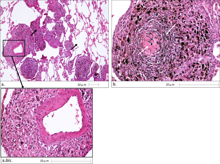
Case presentation: We report the case of a 42-year-old male dental technician who began to suffer from a dry cough and exertional dyspnea after approximately 15 years of work. The operations he conducted for his job resulted in the generation of crystalline silica, aluminum, chromium and titanium dust. The worker did not regularly wear personal protective equipment and some of the above operations were not carried out in closed circuit systems. The Chest X-ray showed diffused micronodules in the pulmonary interstitium of the upper-middle lobes, bilaterally, and a modest left basal pleural effusion. Simple spirometry showed small airway obstruction in its initial stage. High Resolution Computerized Tomography of the chest showed bilateral micronodulation of a miliariform type, with greater profusion to the upper lobes, also present in the visceral pleura, bilaterally. Histological examination showed aggregates of pigment-laden macrophages forming perivascular macules or arranged in a radial pattern around a core of sclerohyalinosis. Scanning Electron Microscopy and Energy Dispersive Spectrometry revealed several mineral particles, typically characterized by the presence of crystalline silica and metal aggregates. The environmental concentrations of total dust and its respirable fraction were all lower than the relative TLV-TWA-ACGIH, yet not negligible.
Conclusions: The above findings and a multidisciplinary assessment led to the diagnosis of mixed dust pneumoconiosis s/q with 2/2 profusion of occupational origin. This diagnosis in a dental technician was supported for the first time in literature by environmental exposure analysis.
Keywords: Dental technician; Environmental analysis; Histologic analysis; Mineralogical analysis; Pneumoconiosis.
© 2022. The Author(s).
Conflict of interest statement
The authors have no conflicts of interest to declare that are relevant to the content of this article.
Figures


References
-
- Quintal-Méndez JR, Soledad AR, López-Hernández E, Sánchez-Monroy V. Pulmonary alterations among workers in a dental prosthesis laboratory: exploring high dust concentrations and novel findings of bacterial genera in the workplace to achieve improved control. J Occup Environ Med. 2020;62:930–936. doi: 10.1097/JOM.0000000000001995. - DOI - PubMed
-
- Romita P, Foti C, Masciopinto L, Nettis E, Di Leo E, Calogiuri G, Bonamonte D, Angelini G, Dipalma G, Ballini A, et al. Allergic contact dermatitis to acrylates. J Biol Regul Homeost Agents. 2017;31:529–534. - PubMed
Publication types
MeSH terms
Substances
LinkOut - more resources
Full Text Sources
