. 2022 Apr 27;20(1):32.
doi: 10.1186/s12969-022-00691-w.

Restricted upper airway dimensions in patients with dentofacial deformity from juvenile idiopathic arthritis

Affiliations

Restricted upper airway dimensions in patients with dentofacial deformity from juvenile idiopathic arthritis

Xiaowen Niu et al. Pediatr Rheumatol Online J. .

Abstract

Background: This retrospective, cross-sectional study aimed to assess the pharyngeal airway dimensions of patients with juvenile idiopathic arthritis (JIA) and moderate/severe JIA-related dentofacial deformity (mandibular retrognathia/micrognathia), and compare the results with JIA patients with a normal mandibular appearance and a group of non-JIA patients.

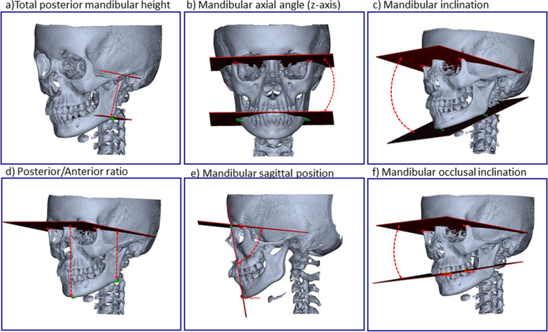
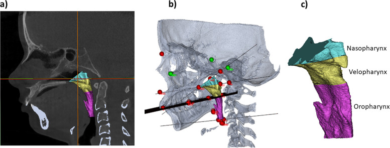
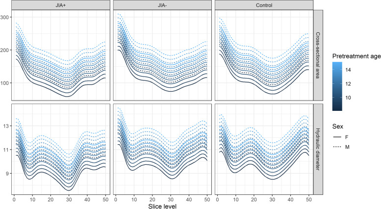
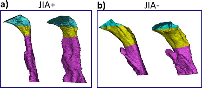
Methods: Seventy-eight patients were retrospectively included in a 1:1:1 manner as specified below. All patients had previously been treated at the Section of Orthodontics, Aarhus University, Denmark. All had a pretreatment cone beam computed tomography (CBCT). Group 1 (JIA+); 26 JIA patients with severe arthritis-related dentofacial deformity and mandibular retrognathia/micrognathia. Group 2 (JIA-); 26 JIA patients with normal mandibular morphology/position. Group 3 (Controls); 26 non-JIA subjects. Dentofacial morphology and upper airway dimensions, excluding the nasal cavity, were assessed in a validated three-dimensional (3D) fashion. Assessment of dentofacial deformity comprised six morphometric measures. Assessment of airway dimensions comprised nine measures.

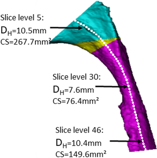
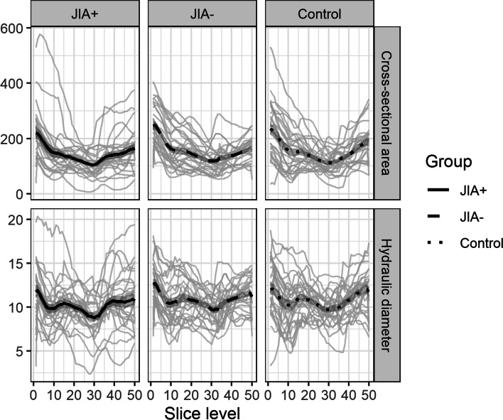
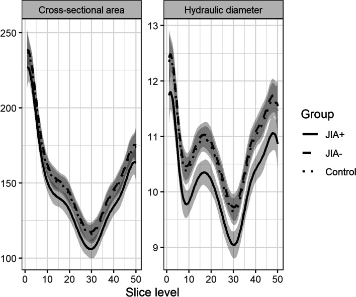
Results: Five morphometric measures of dentofacial deformity were significantly deviating in the JIA+ group compared with the JIA- and control groups: Posterior mandibular height, anterior facial height, mandibular inclination, mandibular occlusal inclination, and mandibular sagittal position. Five of the airway measurements showed significant inter-group differences: JIA+ had a significantly smaller nasopharyngeal airway dimension (ad2-PNS), a smaller velopharyngeal volume, a smaller minimal cross-sectional area and a smaller minimal hydraulic diameter than JIA- and controls. No significant differences in upper airway dimensions were seen between JIA- and controls.

Conclusion: JIA patients with severe arthritis-related dentofacial deformity and mandibular micrognathia had significantly restricted upper airway dimensions compared with JIA patients without dentofacial deformity and controls. The restrictions of upper airway dimension seen in the JIA+ group herein were previously associated with sleep-disordered breathing in the non-JIA background population. Further studies are needed to elucidate the role of dentofacial deformity and restricted airways in the development of sleep-disordered breathing in JIA.


Conflict of interest statement

No competing interests.

Figures

Fig. 1
Fig. 1
Morphometric measures used to assess dentofacial deformity
Fig. 2
Fig. 2
Upper airway dimensions. a and b Upper airways in relation to other anatomical structures. Green and red dots indicate anatomaical landmarks used for evaluation of dentofacial deformity. The black structure indicates the occlusal plane. c Subdivision of upper airways
Fig. 3
Fig. 3
Assessment of cross-sectional area (CS) and hydraulic diameter (DH) along the centerline throughout the upper airway in a patient from the JIA+ group. Each white dot represents a slice level. The middle observation represents the position of the minimum CS and DH (slice level 30). Notice how the upper assessment (slice level 5) and the lower assessment (slice level 46) have comparable DH but great difference in CS due to difference in airway shape
Fig. 4
Fig. 4
Average cross-sectional areas (mm2) and hydraulic diameter (mm) throughout the upper airways from each of the groups. Group 1 (JIA+), group 2 (JIA-), group 3 (controls). Solid black lines represent the mean curve values. The grey lines indicate intra-group curve values for each subject
Fig. 5
Fig. 5
Comparison of cross-sectional areas (mm2) and hydraulic diameter (mm) throughout the upper airways from each of the groups. Group 1 (JIA+), group 2 (JIA-), group 3 (controls)
Fig. 6
Fig. 6
Non-linear mixed effect model combining covariates of sex and pre-treatment age with average cross-sectional area (mm2) and hydraulic diameter (mm) within the three groups. Group 1 (JIA+), group 2 (JIA-), group 3 (controls)
Fig. 7
Fig. 7
An illustration of inter-group differences in upper airway morphology between JIA+ and JIA-. Airways are displayed in lateral and oblique views. a) Upper airway in a patients from the JIA+ group (total volume 8,091.28 mm3). B) Upper airway in a subject from the JIA- group (total volume 8,096.69 mm3). Visual differences are appreciated in sagittal and vertical dimensions of the airways despite comparable total volumes; the JIA+ upper airway presents as a “long and narrow” upper airway

Similar articles

References

    1. Glerup M, Stoustrup P, Matzen LH, Rypdal V, Nordal E, Frid P, et al. Longterm Outcomes of Temporomandibular Joints in Juvenile Idiopathic Arthritis: 17 Years of Followup of a Nordic Juvenile Idiopathic Arthritis Cohort. J Rheumatol. 2020;47(5):730–738. doi: 10.3899/jrheum.190231. - DOI - PubMed
    1. Stoll ML, Sharpe T, Beukelman T, Good J, Young D, Cron RQ. Risk factors for temporomandibular joint arthritis in children with juvenile idiopathic arthritis. J Rheumatol. 2012;39(9):1880–1887. doi: 10.3899/jrheum.111441. - DOI - PubMed
    1. Stoustrup P, Glerup M, Bilgrau AE, Küseler A, Verna C, Christensen AE, et al. Cumulative Incidence of Orofacial Manifestations in Early Juvenile Idiopathic Arthritis: A Regional. Three-Year Cohort Study Arthritis Care Res (Hoboken) 2020;72(7):907–916. doi: 10.1002/acr.23899. - DOI - PubMed
    1. von Schuckmann L, Klotsche J, Suling A, Kahl-Nieke B, Foeldvari I. Temporomandibular joint involvement in patients with juvenile idiopathic arthritis: a retrospective chart review. Scand J Rheumatol. 2020;49(4):271–280. doi: 10.1080/03009742.2020.1720282. - DOI - PubMed
    1. Frid P, Nordal E, Bovis F, Giancane G, Larheim TA, Rygg M, et al. Temporomandibular Joint Involvement in Association With Quality of Life, Disability, and High Disease Activity in Juvenile Idiopathic Arthritis. Arthritis Care Res (Hoboken) 2017;69(5):677–686. doi: 10.1002/acr.23003. - DOI - PubMed

MeSH terms