

Relationship between bone mineral density and ovarian function and thyroid function in perimenopausal women with endometriosis: a prospective study
- PMID: 35477494
- PMCID: PMC9044768
- DOI: 10.1186/s12905-022-01711-3
Relationship between bone mineral density and ovarian function and thyroid function in perimenopausal women with endometriosis: a prospective study
Abstract
Background: In women with endometriosis, the association between ovarian function, hormones, and bone mineral density (BMD) is unclear. Therefore, this study aimed to elucidate the association between changes in bone mineral density (BMD) and clinical data, such as ovarian reserves, in perimenopausal women with endometriosis.
Methods: In this prospective study, we evaluated 207 female patients who visited the Department of Obstetrics and Gynecology at the University of Tokyo Hospital between December 2015 and December 2020. We included patients aged ≥ 40 years with a history of endometriosis or who presented with endometriosis lesions. Patients with a history of smoking, steroid administration, autoimmune diseases, dyslipidaemia, and heart disease were excluded. During the study period, patients who underwent two tests, an initial and a follow-up test (n = 142, average age: 45.02 years, average BMD: 1.16 g/cm2), were evaluated at regular intervals based on the annual rate of change in BMD.
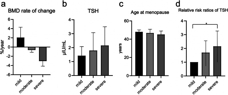
Results: There was a weak negative correlation between the follicle-stimulating hormone (FSH) and BMD and a weak positive correlation between the anti-Müllerian hormone (AMH) and BMD. The annual rate of change in BMD showed a very weak correlation with thyroid-stimulating hormone (TSH) levels. A large decline in BMD was associated with high TSH levels and higher average age at menopause. Patients with higher TSH exhibited a higher rate of decrease in BMD than those without.
Conclusions: High FSH or low AMH levels are associated with decreased BMD. Decreased ovarian reserve is associated with decreased BMD in perimenopausal women with endometriosis. High TSH levels increase the risk of BMD loss. This finding may suggest that women with endometriosis should undergo bone scanning to rule out the possibility of reduced bone mass and subsequent increased risk of fracture.
Keywords: Bone mineral density; Endometriosis; Osteoporosis; Ovarian reserve; Perimenopause.
© 2022. The Author(s).
Conflict of interest statement
The authors declare that they have no competing interests.
Figures
References
Publication types
MeSH terms
Substances
LinkOut - more resources
Full Text Sources
Medical
