. 2022 Aug 3;30(8):2709-2721.
doi: 10.1016/j.ymthe.2022.04.019. Epub 2022 Apr 27.

RNAi-based modulation of IFN-γ signaling in skin

Affiliations

RNAi-based modulation of IFN-γ signaling in skin

Qi Tang et al. Mol Ther. .

Abstract

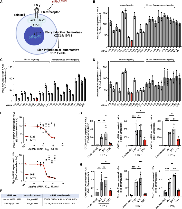
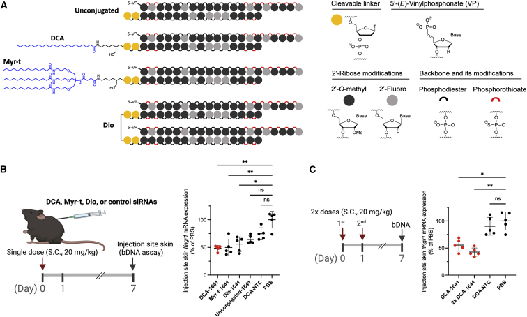
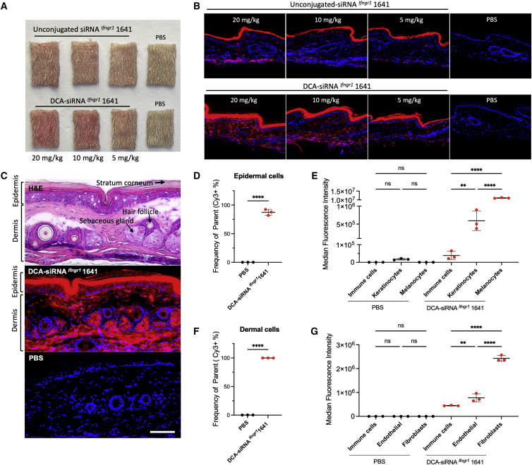
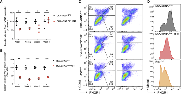
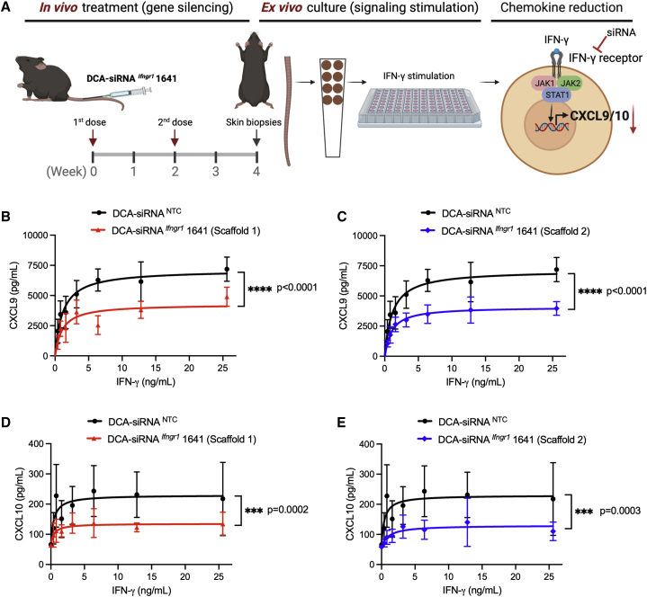
Aberrant activation of interferon (IFN)-γ signaling plays a key role in several autoimmune skin diseases, including lupus erythematosus, alopecia areata, vitiligo, and lichen planus. Here, we identify fully chemically modified small interfering RNAs (siRNAs) that silence the ligand binding chain of the IFN-γ receptor (IFNGR1), for the modulation of IFN-γ signaling. Conjugating these siRNAs to docosanoic acid (DCA) enables productive delivery to all major skin cell types local to the injection site, with a single dose of injection supporting effective IFNGR1 protein reduction for at least 1 month in mice. In an ex vivo model of IFN-γ signaling, DCA-siRNA efficiently inhibits the induction of IFN-γ-inducible chemokines, CXCL9 and CXCL10, in skin biopsies from the injection site. Our data demonstrate that DCA-siRNAs can be engineered for functional gene silencing in skin and establish a path toward siRNA treatment of autoimmune skin diseases.

Keywords: CXCL9/10/11 chemokines; IFN-γ signaling; RNAi therapeutics; autoimmune disorders; immunomodulatory drugs; preclinical drug development; siRNA delivery; skin immunology.


Conflict of interest statement

Declaration of interests A.K., J.E.H., and Q.T. have filed a patent that covers the abovementioned IFNGR1 and other (JAK1, JAK2, and STAT1) identified siRNA oligonucleotides for the modulation of IFN-γ signaling pathway. A.K. discloses ownership of stock in RXi Pharmaceuticals and Advirna and is a founder of Atalanta Therapeutics. J.E.H holds equity in Rheos Medicines, TeVido BioDevices and is a founder of Villaris Therapeutics, Aldena Therapeutics, NIRA Biosciences, and Vimela Therapeutics.

Figures

Figure 1
Figure 1
Identification of human IFNGR1 and mouse Ifngr1 siRNA leads that efficiently modulate IFN-γ signaling (A) Schematic of siRNA silencing IFN-γ receptor for the modulation of IFN-γ signaling. (B) Human IFNGR1 silencing in HeLa cells, (C) mouse Ifngr1 silencing in N2a cells, and (D) human IFNGR1 silencing in SH-SY5Y cells. Cells were treated with fully modified cholesterol-conjugated siRNAs at 1.5 μM for 72 h. The mRNA levels were measured by using QuantiGene 2.0 assays. siRNA number represents the 5′ position of the mRNA target site. UNT, untreated control. Data are represented as percent of UNT (n = 3, mean ± standard deviation). (E) Dose-response curves of lead siRNA compounds (human 1726, mouse 1641). M, molar concentration of siRNA (n = 3, mean ± standard deviation). (F) Targeting region of lead siRNA in human IFNGR1 and mouse Ifngr1 mRNA. (G) Human CXCL9/10/11, and (H) Mouse Cxcl9/10/11 mRNA expressions (QuantiGene 2.0 assay) at 6 h post IFN-γ signaling stimulation; cells were treated with siRNA at 1.5 μM for 72 h before IFN-γ stimulation (n = 4, mean ± standard deviation, one-way ANOVA, ∗p < 0.05, ∗∗p < 0.01, ∗∗∗p < 0.001, ∗∗∗∗p < 0.0001; ns, not significant).
Figure 2
Figure 2
Chemical configurations of siRNA Ifngr1 1641 for gene silencing in mouse skin (A) Schematic of the chemical structures of hydrophobically conjugated (DCA; tri-myristic acid, Myr-t) and Dio siRNAs; DCA and Myr-t conjugates are covalently linked to the 3′ end of sense strand; the two sense strands of the Dio scaffold are covalently linked by a tetraethylene glycol; the study also included unconjugated siRNA Ifngr1 1641 and DCA conjugated NTC siRNAs. (B) Ifngr1 mRNA silencing in skin at the injection site; mice (n = 5 per group) were injected subcutaneously (between shoulders) with a single dose of siRNA (20 mg/kg); local skin was collected at 1 week after injection and mRNA levels were measured using QuantiGene 2.0 assays; Ifngr1 expression was normalized to a housekeeping gene Ppib; data are represented as percent of PBS control (mean ± standard deviation) and analyzed by Kruskal-Wallis test (∗p < 0.05, ∗∗p < 0.01; ns, not significant). (C) Efficacy of a single dose versus two doses (2×, 24 h apart; n = 5) of DCA-siRNA Ifngr1 1641 (One-way ANOVA, ∗p < 0.05, ∗∗p < 0.01). bDNA, branched DNA.
Figure 3
Figure 3
Local distribution profile of DCA-siRNA in skin cell types Single injection of Cy3-labeled siRNA Ifngr1 1641 with or without DCA conjugation (at 3 different doses: 20, 10, 5 mg/kg in 150 μL PBS) were subcutaneously injected into mouse tail skin (a 25G, 40-mm needle was fully inserted and pulled slowly while injecting to cover approximately two-thirds of the area of the tail). Skin samples were collected at 48 h after the injection. (A) Images of skin biopsies from the injection site show local retention of siRNAs (pink), and (B) fluorescence microscopic images show siRNA (red) retention in local skin (nuclei were stained in blue with DAPI, original magnification ×20). (C) Hematoxylin and eosin staining (top) of skin biopsy distinguishes the morphology of epidermis and dermis (transverse section, arrows indicate stratum corneum, sebaceous gland, and hair follicle); Cy3-DCA-siRNA Ifngr1 1641 distribution (red in middle panel); PBS background staining (bottom panel); original magnification ×20, bar scale = 100 μm. (D) Percentage of Cy3-positive cell population in live epidermal cells (n = 3, mean ± standard deviation, 20 mg/kg DCA-siRNA Ifngr1 1641, and PBS background control, unpaired t-test, ∗∗∗∗p < 0.0001), and (E) cell size-normalized median fluorescence intensity (MFI) indicates relative delivery efficiency of Cy3-siRNA in epidermal cell types (one-way ANOVA for multiple comparison; ∗p < 0.05, ∗∗∗∗p < 0.0001; ns, not significant). (F) Percentage of Cy3 positive cell population in live dermal cells, and (G) normalized MFI in dermal cell types (∗∗∗p < 0.001, ∗∗∗∗p < 0.0001).
Figure 4
Figure 4
Duration of Ifngr1 silencing by siRNA Ifngr1 1641 in mouse tail skin (A) Ifngr1 mRNA levels over four weeks post injection (n = 3 per group, single dose at 20 mg/kg); mRNA levels were measured using QuantiGene 2.0 assays, Ifngr1 expression was normalized to the housekeeping gene Ppib; data were represented as percent of NTC control (mean ± standard deviation) and analyzed by unpaired t test (∗p < 0.05; ns, not significant). (B) IFNGR1 protein levels were determined by the relative MFI of anti-mouse IFNGR1 staining in CD45+ (hematopoietic cell marker) cell population; MFI values were subtracted from Ifngr1−/− background staining, and normalized to NTC control (mean ± standard deviation, unpaired t test, ∗∗p < 0.01, ∗∗∗p < 0.001). (C) Representative flow cytometry panels of CD45+ cells and IFNGR1 staining at week 2 post-injection. (D) Histograms of graph (C) for IFNGR1 staining in CD45+ cells.
Figure 5
Figure 5
DCA-siRNA Ifngr1 1641 inhibits IFN-γ signaling in an ex vivo skin model (A) Schematic of siRNA treatment in mice and stimulation of IFN-γ signaling in an ex vivo skin biopsy model; mice (n = 5 per group) were treated subcutaneously with two doses (2×; 20 mg/kg; 2 weeks apart) of DCA-siRNA Ifngr1 1641 in tail skin, and eight punches per mouse of skin biopsies (4 mm in diameter) were collected. For each mouse, a seven-point dose response of IFN-γ signaling stimulation was carried out using recombinant mouse IFN-γ at a concentration range of 0–25.6 ng/mL. Skin biopsies were incubated at 37°C for 24 h, and CXCL9 and CXCL10 levels were determined by ELISA. (B–E) Production of CXCL9 and CXCL10 after the treatment of two scaffold configurations of DCA-siRNA Ifngr1 1641 (two-way ANOVA, ∗∗∗p < 0.001, ∗∗∗∗p < 0.0001).

References

    1. Ruffilli I. MIG in cutaneous systemic lupus erythematosus. Clin. Ter. 2019;170:e71–e76. doi: 10.7417/CT.2019.2111. - DOI - PubMed
    1. Rork J.F., Rashighi M., Harris J.E. Understanding autoimmunity of vitiligo and alopecia areata. Curr. Opin. Pediatr. 2016;28:463–469. doi: 10.1097/mop.0000000000000375. - DOI - PMC - PubMed
    1. Harris J.E., Harris T.H., Weninger W., John Wherry E., Hunter C.A., Turka L.A. A mouse model of vitiligo with focused epidermal depigmentation requires IFN-γ for autoreactive CD8+ T-cell accumulation in the skin. J. Invest. Dermatol. 2012;132:1869–1876. doi: 10.1038/jid.2011.463. - DOI - PMC - PubMed
    1. Shao S., Tsoi L.C., Sarkar M.K., Xing X., Xue K., Uppala R., Berthier C.C., Zeng C., Patrick M., Billi A.C., et al. IFN-γ enhances cell-mediated cytotoxicity against keratinocytes via JAK2/STAT1 in lichen planus. Sci. Transl. Med. 2019;11:eaav7561. doi: 10.1126/scitranslmed.aav7561. - DOI - PMC - PubMed
    1. Rashighi M., Agarwal P., Richmond J.M., Harris T.H., Dresser K., Su M.-W., Zhou Y., Deng A., Hunter C.A., Luster A.D., et al. CXCL10 is critical for the progression and maintenance of depigmentation in a mouse model of vitiligo. Sci. Transl. Med. 2014;6:223ra23. doi: 10.1126/scitranslmed.3007811. - DOI - PMC - PubMed

Publication types