. 2022 Jun 1;42(22):4517-4537.
doi: 10.1523/JNEUROSCI.1786-21.2022. Epub 2022 Apr 27.

Human Spindle Variability

Affiliations

Human Spindle Variability

Christopher Gonzalez et al. J Neurosci. .

Abstract

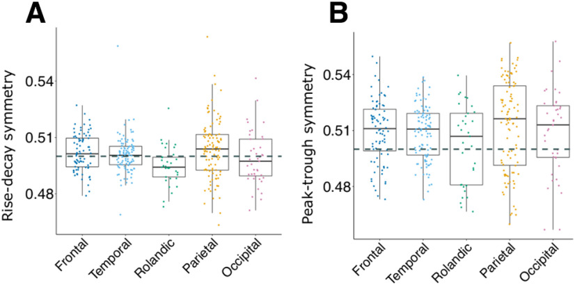
In humans, sleep spindles are 10- to 16-Hz oscillations lasting approximately 0.5-2 s. Spindles, along with cortical slow oscillations, may facilitate memory consolidation by enabling synaptic plasticity. Early recordings of spindles at the scalp found anterior channels had overall slower frequency than central-posterior channels. This robust, topographical finding led to dichotomizing spindles as "slow" versus "fast," modeled as two distinct spindle generators in frontal versus posterior cortex. Using a large dataset of intracranial stereoelectroencephalographic (sEEG) recordings from 20 patients (13 female, 7 male) and 365 bipolar recordings, we show that the difference in spindle frequency between frontal and parietal channels is comparable to the variability in spindle frequency within the course of individual spindles, across different spindles recorded by a given site, and across sites within a given region. Thus, fast and slow spindles only capture average differences that obscure a much larger underlying overlap in frequency. Furthermore, differences in mean frequency are only one of several ways that spindles differ. For example, compared with parietal, frontal spindles are smaller, tend to occur after parietal when both are engaged, and show a larger decrease in frequency within-spindles. However, frontal and parietal spindles are similar in being longer, less variable, and more widespread than occipital, temporal, and Rolandic spindles. These characteristics are accentuated in spindles which are highly phase-locked to posterior hippocampal spindles. We propose that rather than a strict parietal-fast/frontal-slow dichotomy, spindles differ continuously and quasi-independently in multiple dimensions, with variability due about equally to within-spindle, within-region, and between-region factors.SIGNIFICANCE STATEMENT Sleep spindles are 10- to 16-Hz neural oscillations generated by cortico-thalamic circuits that promote memory consolidation. Spindles are often dichotomized into slow-anterior and fast-posterior categories for cognitive and clinical studies. Here, we show that the anterior-posterior difference in spindle frequency is comparable to that observed between different cycles of individual spindles, between spindles from a given site, or from different sites within a region. Further, we show that spindles vary on other dimensions such as duration, amplitude, spread, primacy and consistency, and that these multiple dimensions vary continuously and largely independently across cortical regions. These findings suggest that multiple continuous variables rather than a strict frequency dichotomy may be more useful biomarkers for memory consolidation or psychiatric disorders.

Keywords: cortex; hippocampus; intracranial; sleep; slow oscillation; spindle.


Figures

Figure 1.
Figure 1.
Sleep spindles recorded from human cortex. The spatial locations of six cortical recordings from one patient are shown in A. The average PSDs during spindles for this patient, split by all frontal (n=5) and parietal (n=7) channels, are shown in B. Solid black lines mark the spindle band, 10 and 16 Hz, and the dashed blue line 12 Hz. An example epoch where all six channels are simultaneously spindling is shown in C, with black indicating the LFP, red detected spindles, and blue the bandpass filtered signal in the 10- to 16-Hz range. Colored dots correspond to the spatial locations in A. The average time-frequency plots are shown in D for each cortical channel, with time 0 indicating the deepest trough in the 10- to 16-Hz bandpass. Overlaid on top of the time-frequency plots are the average LFP for each channel. The bottom left of each panel in C indicates the average overall frequency for each channel, and the bottom right the total number of spindles for that channel analyzed. E, Example spindle for the superior frontal channel in panel A. The red lines indicate the start and stop time of the automatic spindle detection, which corresponds to our duration measure. The enlarged spindle panel shows a 10- to 16-Hz signal in blue overlaid, with peaks in the filtered signal marked as dots. Cycles that were of sufficient amplitude in the filtered signal (exceeding 30% of the largest cycle) are marked as green and used for further analysis. For each spindle, the surviving cycles were used to calculate the overall spindle frequency, variation in frequency across cycles, and to estimate frequency change (lower right panel of E). Each panel of F indicates the distribution across all spindles for the same superior frontal channel for multiple spindle features.
Figure 2.
Figure 2.
Primary spindle characteristics. Cortical bipolar sEEG recordings denoted as circles overlaid on an average surface, with warmer colors indicating greater values. Also shown at the top of the brain surfaces for each measure are boxplots grouped by brain region. For the boxplots, different colors and columns denote different regions, box margins indicate interquartile ranges, and dots indicate individual cortical channels. A, Average overall frequency at individual sites is shown. Rolandic (β = 0.41 Hz, t = 3.93) and parietal cortex (0.71 Hz, t = 8.28) show faster overall frequency than frontal cortex. B, Average spindle duration across spindles at individual sites is shown. Temporal (β = −0.03 s, t = −5.52) and occipital (β = −0.02 s, t = −3.03) cortex show shorter duration spindles compared with frontal. C, Average spindle trough-to-peak amplitude is significantly greater in parietal (β = 30.48 µV, t = 2.72) and occipital cortex occipital (β = 62.48 µV, t = 4.52) than frontal cortex. D, Number of spindles per minute during stage 2 (N2); temporal (N2: β = −1.4 spindles/min, t = −6.45) and occipital cortex (N2: β = −1.21 spindles/min, t = −4.37) had lower spindle densities than frontal cortex.
Figure 3.
Figure 3.
Waveform shape across regions. A, Average rise-decay symmetry for each bipolar recording (indicated as dots), divided by cortical region. B, Same as A, for peak-trough symmetry. Dashed lines indicate 0.5. Overall, cortical sites were symmetrical in rise-decay symmetry (β = 2e-03, t = 1.48), and showed a slight but statistically significant bias for longer peaks than troughs (β = 8e-03, t = 2.92).
Figure 4.
Figure 4.
Spindle PSDs for frontal and parietal cortex. Six patients had at least three frontal and three parietal channels, and the average PSD for all channels for each region are shown in A. These PSDs have been corrected by removing each channel's aperiodic, 1/f trend. Average PSDs are primarily unimodal; however, patients 1, 5, 12, and 16 show quite broad PSDs for parietal cortex. B, PSDs as colormaps with all cortical channels on the y-axis, gray lines separating unique patients, and frequency on the x-axis. Again, power has been corrected by removing the aperiodic component. Channels from patients in A are labeled. This display allows the inspection of each channel's peak frequency and the number of peaks for each channel. The majority of channels appear to show a single peak, and some channels show quite broad peaks (e.g., pt 1).
Figure 5.
Figure 5.
Sources of frequency variability. Cortical bipolar sEEG recordings denoted as circles overlaid on an average surface, with warmer colors indicating greater values. Black triangles mark 0.64 Hz (or Hz/s) to indicate the average difference between frontal and parietal sites. Also shown at the top of the brain surfaces for each measure are boxplots grouped by brain region. A, At individual cortical sites, the SD of overall frequency across spindles in temporal (β = 0.07, t = 2.92), Rolandic (β = 0.20, t = 6.46), parietal (β = 0.11, t = 4.27), and occipital cortex (β = 0.17, t = 5.41) showed greater interspindle variability than frontal cortex. B, The average SD across cycle frequency within a spindle. Temporal (β = 0.14 Hz, t = 5.98) and occipital (β = 0.1, t = 3.6) cortex showed greater intraspindle SD compared with frontal cortex. C, Another measure of intraspindle frequency variation, the average difference between the fastest and slowest cycle within a spindle, is 2.7 Hz on average. D, Average estimated linear change in frequency within a spindle (cyan indicates spindle slowing, pink spindle speeding). Frontal cortex showed significantly more intraspindle slowing than temporal (β = 0.64 Hz/s, t = 9.32), Rolandic (β = 0.35 Hz/s, t = 3.98), parietal (β = 0.47, t = 6.66), and occipital cortex (β = 0.60 Hz/s, t = 6.9). The amount of inter and intraspindle variability often exceeds the observed frontal-parietal overall difference in frequency.
Figure 6.
Figure 6.
Spindle co-occurrences across the cortex. A, D, Brain surfaces overlaid with cortical sEEG recording sites. Warmer colors indicate greater values. A, The average proportion of channels participating in a spindle at each cortical site; frontal and parietal show the greatest proportion. B, Distribution of the proportion of channels participating in a spindle (top) and absolute number of channels participating in a spindle (bottom) for all spindles from all channels. C, For each region, the proportion of spindles that occurred in one to eight channels. D, For each channel, the proportion of times a spindle initiated a co-spindling event, i.e., when a spindle co-occurred in at least two channels.
Figure 7.
Figure 7.
Cortical-hippocampal spindle phase locking. A, A cortical precuneus channel from one patient with significant phase-locking during spindles with the posterior hippocampus during N3; time 0 indicates the onset of spindles in the hippocampus. PLV increases after spindle onset and peaks around 0.4 at 750 ms. B, Channels with significant PLV during spindles with a posterior hippocampal channel during N2, as identified by Jiang et al. (2019b), are indicated as circles, with the peak PLV shown in color. Nonsignificant channels are displayed as crosses. Statistical relationships between PLV in significant channels and spindle features are reported in Table 9 and Extended Data Table 9-1. Areas with the highest PLV are apparent in parietal cortex as well as posterior ventral temporal cortex. We performed post hoc analyses comparing these high PLV channels (PLV >0.4) and all other cortical channels, including those with nonsignificant PLV, across several spindle characteristics. Differences between high and low PLV channels in spindle features are reported in Table 10 and Extended Data Table 10-1. C–F, Each dot is a channel, different colors denote different brain regions. High PLV channels were significantly faster (LMEM; β = 0.57, t = 5.01, number of channels = 341, N = 20), (D) had lower intraspindle frequency variation (β = −0.15, t = −4.7), (E) had slightly longer duration (β = 0.02, t = 2.82), and (F) had greater spindle density in N2 (β = 0.76, t = 2.48). These results did not change when restricting to just parietal channels.
Figure 8.
Figure 8.
Times of spindles relative to downstate troughs. The polarity of each cortical bipolar recording is corrected such that negative indicates the trough of the DS, as determined by suppression of high γ (70–190 Hz), shown in A as the grand average across all cortical channels. B, An example epoch where spindles are coupled to downstates for a superior parietal channel from one patient. C, For the same channel in B, a histogram of the occurrence of spindles starting around the DS trough. The probability of spindles starting relative to downstates for all channels with spindles that significantly started before or after downstate troughs are shown in D, E. In D, each gray line corresponds to a bipolar, cortical recording, with black lines indicating average across channels and blue lines chance occurrence. E, Each row is a cortical channel, with gray lines separating different patients and magenta lines indicating the time of downstate trough. The y-axis in D and color in E indicate the probability of spindles starting in a particular time bin. For each region, all spindles starting within ±1 s of the downstate trough are included. The most consistent effect across channels is that spindles have the highest probability of starting after the downstate trough, most apparent in frontal and parietal cortex.
Figure 9.
Figure 9.
Slower and faster spindles more likely to follow than precede downstates. A, B, Cortical channels with at least 100 spindles < 12 Hz and 100 spindles > 12 Hz starting within ±1 s of downstate trough are shown. Rows indicate cortical channels, gray lines separate patients, and the dashed magenta line indicates time of the downstate trough. Color indicates the probability of spindles starting within that channel. For both regions and frequency groups, there is a greater probability of spindles starting after the downstate trough compared with before. This is statistically assessed in C, D, where LMEMs, with patient as random effect, modeled the probability of spindles starting at each time bin for each frequency condition separately, with pink triangles showing spindles >12 Hz and gray squares for spindles <12 Hz. Error bars reflect SE, and the blue dashed line indicates the probability of spindles starting by chance. Time bins where error bars do not include the chance line indicate significance at p < 0.05. Overall, spindles were more likely to start after the downstate trough than before. The degree to which spindles preferentially occur after the downstate is less for spindles <12 Hz.

References

    1. Ameijeiras-Alonso J, Crujeiras RM, Rodriguez-Casal A (2021) multimode: An R Package for Mode Assessment. J Stat Softw 97:1–32.
    1. Amunts K, Zilles K (2015) Architectonic mapping of the human brain beyond Brodmann. Neuron 88:1086–1107. 10.1016/j.neuron.2015.12.001 - DOI - PubMed
    1. Anderer P, Klösch G, Gruber G, Trenker E, Pascual-Marqui RD, Zeitlhofer J, Barbanoj MJ, Rappelsberger P, Saletu B (2001) Low-resolution brain electromagnetic tomography revealed simultaneously active frontal and parietal sleep spindle sources in the human cortex. Neuroscience 103:581–592. 10.1016/S0306-4522(01)00028-8 - DOI - PubMed
    1. Andrillon T, Nir Y, Staba RJ, Ferrarelli F, Cirelli C, Tononi G, Fried I (2011) Sleep spindles in humans: insights from intracranial EEG and unit recordings. J Neurosci 31:17821–17834. 10.1523/JNEUROSCI.2604-11.2011 - DOI - PMC - PubMed
    1. Ayoub A, Aumann D, Hörschelmann A, Kouchekmanesch A, Paul P, Born J, Marshall L (2013) Differential effects on fast and slow spindle activity, and the sleep slow oscillation in humans with carbamazepine and flunarizine to antagonize voltage-dependent Na+ and Ca2+ channel activity. Sleep 36:905–911. 10.5665/sleep.2722 - DOI - PMC - PubMed

Publication types