. 2022 Oct;29(10):2060-2069.
doi: 10.1038/s41418-022-00997-y. Epub 2022 Apr 27.

GSK3β mediates the spatiotemporal dynamics of NLRP3 inflammasome activation

Affiliations

GSK3β mediates the spatiotemporal dynamics of NLRP3 inflammasome activation

Suyavaran Arumugam et al. Cell Death Differ. 2022 Oct.

Abstract

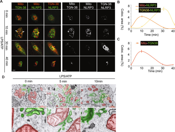
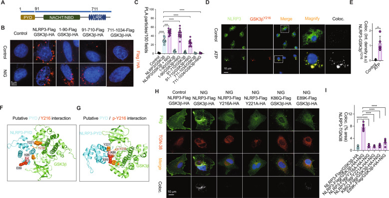
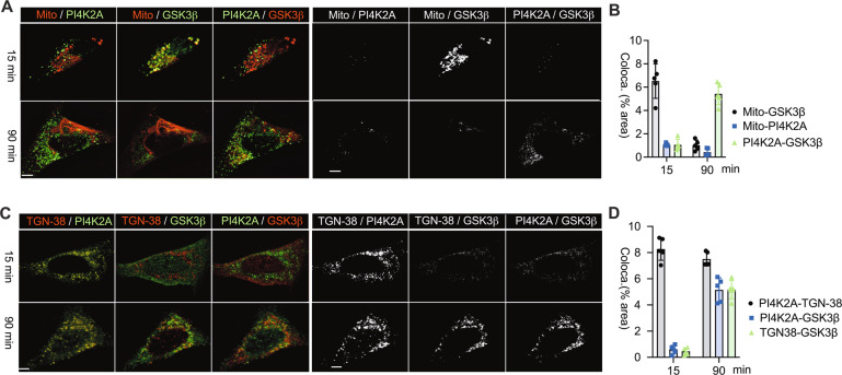
Subcellular machinery of NLRP3 is essential for inflammasome assembly and activation. However, the stepwise process and mechanistic basis of NLRP3 engagement with organelles remain unclear. Herein, we demonstrated glycogen synthase kinase 3β (GSK3β) as a molecular determinant for the spatiotemporal dynamics of NLRP3 inflammasome activation. Using live cell multispectral time-lapse tracking acquisition, we observed that upon stimuli NLRP3 was transiently associated with mitochondria and subsequently recruited to the Golgi network (TGN) where it was retained for inflammasome assembly. This occurred in relation to the temporal contact of mitochondria to Golgi apparatus. NLRP3 stimuli initiate GSK3β activation with subsequent binding to NLRP3, facilitating NLRP3 recruitment to mitochondria and transition to TGN. GSK3β activation also phosphorylates phosphatidylinositol 4-kinase 2 Α (PI4k2A) in TGN to promote sustained NLRP3 oligomerization. Our study has identified the interplay between GSK3β signaling and the organelles dynamics of NLRP3 required for inflammasome activation and opens new avenues for therapeutic intervention.


Conflict of interest statement

The authors declare no competing interests.

Figures

Fig. 1
Fig. 1. Sequential localization of NLRP3 to the mitochondria, association of mitochondria with TGN, and NLRP3 recruitment to TGN.
HeLa cells expressing fluorescent fusion proteins NLRP3-GFP (green), TOM20-mPLUM (red) and Golgi-RFP (CellLightTM) (yellow) were stimulated with nigericin. The time-lapse imaging of live cells was captured every 5 min over the course of 120 min. A Representative snapshot images of NLRP3 dynamic contact with mitochondria and Golgi, and mitochondrial dynamic contact with Golgi were determined by fluorescence merge and colocalization analysis. All the panels represent images acquired from the same cell at various time points. The white pixel intensities show corresponding Z-slices with optimal colocalization over 2 channels obtained from Imaris 9.2.1- colocalization plugin. Scale bar 10 µm. B, C The levels of colocalized pixels between NLRP3 and mitochondria, NLRP3 and Golgi (B), and mitochondria and Golgi vesicles (C) were quantified using Image J. Three biological replicates were analyzed. The error bar indicates SEM. D Time-lapse tracking analysis of NLRP3 dynamic contact with mitochondria and Golgi, and mitochondrial dynamic contact with Golgi. E, F Immunoblot analysis of dynamic NLRP3 protein levels in Trans- (E) and Cis- (F) Golgi fractions that were fractionated from Hela cells expressing NLRP3 with nigericin stimulus as indicated time-course. Purity of the fractions was assessed by blotting for GM130 (Cis-), TGN38 (Trans-), and Tom20 (mitochondria).
Fig. 2
Fig. 2. Subcellular trafficking of NLRP3 in primary mouse BMDMs upon stimulus.
A Representative confocal images of mitochondria-TGN38, mitochondria-NLRP3 and NLRP3-TGN38 contact during 0, 10, 30 and 40 min after LPS/ATP in primary mouse BMDMs. Each 2-channel interaction was false colored and shown as red-green combo. The right panel shows corresponding colocalization pixels of the images on left panel as quantified using Image J. B Quantification of mitochondria-NLRP3 and TGN38-NLRP3 colocalization. C Quantification of mitochondria-TGN38 colocalization. D Electron micrograph shows dynamic contact between mitochondria and Golgi during 0, 5, and 10 min after LPS/ATP stimulation in primary mouse BMDMs. The TEM images were false colored to highlight mitochondria (red) and Golgi structures (green). At least 8 cells were randomly imaged per group per experiment. Scale bar = 10 μm. Data represent the mean +/- SD of three independent experiments.
Fig. 3
Fig. 3. GSK3β deletion blocks NLRP3 trafficking to mitochondria following Golgi upon stimulus.
Hela cells expressing NLRP3-GFP were subjected to transfection with GSK3β specific siRNA (GSK3B KD) and scramble control siRNA (SCR). Cells were stimulated with nigericin (10 µM) for 120 min. A Representative time-lapse snapshots of NLRP3-Golgi and NLRP3-mitochondria contact. Scale bar = 10 µm. (B) Quantification of NLRP3-Mitochondria and NLRP3-Golgi interaction over the time-course in both SCR and GSK3B KD cells. Data represent the mean ± SD of at least three independent experiments.
Fig. 4
Fig. 4. GSK3β is required for NLRP3 trafficking and inflammasome activation.
A Hela cells expressing NLRP3-GFP were treated with GSK3β inhibitor (SB216763) or an antioxidant (N-acetylcysteine (NAC)) following nigericin stimulation over 120 min. Representative images of NLRP3 aggregation on Trans-Golgi determined by fluorescence merge and colocalization analysis. B The levels of NLRP3- TGN38 colocalized pixel intensities were quantified from 10 different fields in each group. ****P < 0.0001. CG LPS-primed mouse BMDMs were treated with SB216763 at different dosages following ATP (5 mM) for 30 min (C, EG), or nigericin (10 µM) for 40 min (D). C, D Immunoblot analysis of active caspase-1 and IL-1β release, NLRP3 protein content and GSK3β phosphorylation were measured by western blot. E IL-1β release in culture supernatants by ELISA. F Immunofluorescence imaging of ASC specks (green spots indicated with white arrowheads) was performed using the antibody against ASC. Nuclei were stained with DAPI (blue). G The levels of ASC specks were quantified by Image J. n = 12 fields. *P < 0.01. H IL-1β release by ELISA in culture supernatants from LPS-primed Raw264.7 macrophages stably expressing ASC with treatment as described in C. I IL-1β release by ELISA in culture supernatants from LPS-primed J774 macrophages which were pretreated with SB216763 following ATP as described in C. Data represent mean ± SD of three independent experiments. *P < 0.05, **P < 0.01.
Fig. 5
Fig. 5. pGSK3β-Tyr216 interacts with NLRP3 via its pyrin domain.
A Diagram of NLRP3 fragment constructs containing PYD (1-90), NBD (91-710), and LRR (711-1034) domain. B Representative images of Duolink proximity ligation (PLA) assay of Hela cells in situ co-transfected with GSK3β-HA tag and NLRP3-Flag tag fragment constructs with/without nigericin stimulation. C The levels of PLA fluorescence pixels quantified by Image J. n = 10 fields. Scale bar 10 µm. ****P < 0.001, ***P < 0.01, **P < 0.05. Representative images (D) and quantifications (E) of NLRP3 colocalization with phosphorylated GSK3β at Tyr216 site in LPS-primed BMDMs as described in Fig. 2C. n = 6 fields. Scale bar 10 µm. Data represent mean ± SD of three independent experiments. *P < 0.05. F, G Putative interaction between NLRP3PYD domain (PDB ID: 2NAQ) and GSK3β (PDB ID: 4DIT) with phosphorylation at Y216 site was predicated using HADDOCK protein-protein docking program by Haddock prediction interface. F, G Pymol generated images of docking results show a strong interaction between (F) K86 of NLRP3PYD and Y221 of GSK3β while the mutation of GSK3β-Y221A results in a weak interaction with K86 as shown in (G). H, I NLRP3-TGN38 colocalization was disrupted by the co-expression of NLRP3PYD/ GSK3β-Y216A, NLRP3PYD/ GSK3β-Y221A, NLRP3PYD-K86Q/ GSK3β, or NLRP3PYD-E89K mutant constructs, (H) Representative images, (I) Quantifications. n = 10 fields. Scale bar 10 µm. Data represent mean ± SD of three independent experiments. ****P < 0.001, ***P < 0.01.
Fig. 6
Fig. 6. GSK3β directly targets on PI4K2A at TGN upon stimulus.
Hela cells were stimulated with nigericin (10 μM) for 15 and 90 min. A, B Representative images and the quantification of mitochondria/PI4K2A, mitochondria/GSK3β, PI4K2A/GSK3β colocalization. C, D Representative images and the quantification of TGN-38/PI4K2A, TGN-38/GSK3β, PI4K2A/ GSK3β colocalization. Scale bar = 10 μm. The 2 channel colocalization was quantified using Image J colocalization plugin. At least 5 cells were imaged per group and quantified. Data represent the mean +/- SD of three independent experiments.
Fig. 7
Fig. 7. PI4K2A phosphorylation by GSK3β directs NLRP3 recruitment to the TGN.
A Representative images of Duolink-PLA of Hela cells co-transfected with PI4K2A-His and NLRP3-Flag constructs. B The levels of PLA fluorescence pixels were quantified by Image J. n = 10 fields. Scale bar 10 µM. ****P < 0.001, ***P < 0.01, **P < 0.05. C Representative images of NLRP3 colocalization with PI4k2A in LPS/Nigericin treated BMDMs. D Mass spectrometry identification of highly conserved PI4k2A peptides in phosphorylated proteins enriched from the cell lysate of LPS/ATP treated BMDMs. E Representative images and (F) quantification of NLRP3 colocalization with Trans-Golgi in nigericin stimulated Hela cells expressing PI4K2A-S5/9A mutant. n = 8 fields. Scale bar 10 µM. ***P < 0.01. G Endogenous GSK3β association with PI4k2A and NLRP3 in LPS/ATP treated BMDMs. H Immunoblot analysis of active caspase-1 and IL-1β release, the protein contents of NLRP3, GSK3β and PI4k2A in whole cell lysate (Lysate) and cell supernatants (Super) from LPS/ATP treated BMDMs with Gsk3β and Pi4k2α knockdown (KD). I IL-1β release by ELISA in cell supernatants from LPS/ATP treated BMDMs with Gsk3β and Pi4k2α knockdown (KD). Data represent mean ± SD of three independent experiments. *P < 0.05, **P < 0.01.

References

    1. Place DE, Kanneganti TD. Recent advances in inflammasome biology. Curr Opin Immunol. 2018;50:32–8. doi: 10.1016/j.coi.2017.10.011. - DOI - PMC - PubMed
    1. Guo H, Callaway JB, Ting JP. Inflammasomes: Mechanism of action, role in disease, and therapeutics. Nat Med. 2015;21:677–87. doi: 10.1038/nm.3893. - DOI - PMC - PubMed
    1. Lu A, Magupalli VG, Ruan J, Yin Q, Atianand MK, Vos MR, et al. Unified polymerization mechanism for the assembly of ASC-dependent inflammasomes. Cell. 2014;156:1193–206. doi: 10.1016/j.cell.2014.02.008. - DOI - PMC - PubMed
    1. Cai X, Chen J, Xu H, Liu S, Jiang QX, Halfmann R, et al. Prion-like polymerization underlies signal transduction in antiviral immune defense and inflammasome activation. Cell. 2014;156:1207–22. doi: 10.1016/j.cell.2014.01.063. - DOI - PMC - PubMed
    1. Chen J, Chen ZJ. PtdIns4P on dispersed trans-Golgi network mediates NLRP3 inflammasome activation. Nature. 2018;564:71–6. doi: 10.1038/s41586-018-0761-3. - DOI - PMC - PubMed

Publication types

MeSH terms

Substances