. 2022 Apr;11(2):e1270.
doi: 10.1002/mbo3.1270.

Recombinant Limosilactobacillus (Lactobacillus) delivering nanobodies against Clostridium perfringens NetB and alpha toxin confers potential protection from necrotic enteritis

Affiliations

Recombinant Limosilactobacillus (Lactobacillus) delivering nanobodies against Clostridium perfringens NetB and alpha toxin confers potential protection from necrotic enteritis

Dharanesh Gangaiah et al. Microbiologyopen. 2022 Apr.

Abstract

Necrotic enteritis (NE), caused by Clostridium perfringens, is an intestinal disease with devastating economic losses to the poultry industry. NE is a complex disease and predisposing factors that compromise gut integrity are required to facilitate C. perfringens proliferation and toxin production. NE is also characterized by drastic shifts in gut microbiota; C. perfringens is negatively correlated with Lactobacilli. Vaccines are only partially effective against NE and antibiotics suffer from the concern of resistance development. These strategies address only some aspects of NE pathogenesis. Thus, there is an urgent need for alternative strategies that address multiple aspects of NE biology. Here, we developed Limosilactobacillus (Lactobacillus) reuteri vectors for in situ delivery of nanobodies against NetB and α toxin, two key toxins associated with NE pathophysiology. We generated nanobodies and showed that these nanobodies neutralize NetB and α toxin. We selected L. reuteri vector strains with intrinsic benefits and demonstrated that these strains inhibit C. perfringens and secrete over 130 metabolites, some of which play a key role in maintaining gut health. Recombinant L. reuteri strains efficiently secreted nanobodies and these nanobodies neutralized NetB. The recombinant strains were genetically and phenotypically stable over 480 generations and showed persistent colonization in chickens. A two-dose in ovo and drinking water administration of recombinant L. reuteri strains protected chickens from NE-associated mortality. These results provide proof-of-concept data for using L. reuteri as a live vector for delivery of nanobodies with broad applicability to other targets and highlight the potential synergistic effects of vector strains and nanobodies for addressing complex diseases such as NE.

Keywords: Limosilactobacillus; nanobodies; necrotic enteritis; poultry.


Conflict of interest statement

Dharanesh Gangaiah, Valerie Ryan, Shrinivasrao P. Mane, Enid T. McKinley, Nandakumar D. Reddy, and Arvind Kumar are employees of Elanco Animal Health Inc. Nallakannu Lakshmanan was an employee of Elanco Animal Health Inc. at the time the work was done. Elanco Animal Health Inc. is a company that develops, manufactures, and sells veterinary pharmaceuticals and nutritionals. Daphne van Hoese and Edward Dolk are current employees of QVQ Holding BV, which discovers and develops nanobodies for different target diseases.

Figures

Figure 1
Figure 1
Llama immunization scheme (a) and immune response of SNL‐133 (b and d) and SNL‐134 (c and e) to directly coated α toxin (b and c) and NetB (d and e). Two llamas, SNL133 and SNL134, were immunized with NetB and α toxin toxoids on Day 0, Day 14, Day 28, Day 35, Day 57, and Day 71, and sera were collected at different time points and analyzed for an immune response using ELISA. Representative results are shown from three independent experiments
Figure 2
Figure 2
SDS‐PAGE analysis of the purified VHHs and their dose response binding to NetB and α toxin. (a) Coomassie‐stained SDS‐PAGE showing analysis of purified VHH selected on NetB (1–5) and α toxin (6–12). R, 1 µg reference VHH; L, Prestained protein ladder (PageRuler, ThermoFisher); 1, ENB‐1A4; 2, ENB‐1F4; 3, ENB‐1B9; 4, ENB‐1F10; 5, ENB‐1D11; 6, EAT‐1A2; 7, EAT‐1F2; 8, EAT‐1G4; 9, EAT‐1F3; 10, EAT‐1D7; 11, EAT‐1A3; and 12, EAT‐1C8. (b) and (c) Dose response binding of the selected VHHs to α toxin (b) or NetB (c). Representative results are shown from three independent experiments
Figure 3
Figure 3
Neutralization of α toxin lecithinase and NetB hemolytic activity by VHH clones. (a, b) A twofold dilution series of VHH antibodies, starting from 5 µM concentration was preincubated with either (a) recombinant α toxin or (b) commercial α toxin, after which egg yolk solution was added. Serum derived from calves immunized with recombinant α toxin was used as a control (control serum). Egg yolk solution incubated with α toxin without VHH, or serum was used to calculate 100% activity. (c) A twofold dilution series of VHH, starting from 5 µM concentration, was preincubated with recombinant NetB (in a total volume of 2 µl), after which 1% chicken erythrocytes was added. Serum derived from rabbits immunized with recombinant NetB was used as a control (control serum). The optical density of 100% hemolysis (mean OD570 = 0.37, indicated by solid line) was obtained by diluting the chicken erythrocytes in distilled water. As a control, chicken erythrocytes incubated with NetB, but without VHH or serum was used (mean OD570 = 0.36, indicated by dotted line). This resulted in 100% hemolysis. A phosphate‐buffered saline control (1% chicken erythrocytes with no NetB or Nbs) resulted in a mean OD570 of 0.03. Representative results are shown from three independent experiments. VHH, Variable domain of the Heavy chain of Heavy chain
Figure 4
Figure 4
Optimization of VHH clones for improved affinity, production, and stability. (a) Structure of ENB‐ID11 predicted based on homology modeling. The (b, c) Affinity of VHH clones to α toxin (EAT clones) and NetB (ENB clones). (dg) SDS‐PAGE gel of stable VHH control (d), EGFR Q44C (protease susceptible control) (e), EAT‐1F2 (f), and ENB‐1D11_R56H (g) incubated with immobilized trypsin for different time points: 0, 15, 30, 45, 60, 90, and 120 min at 37°C. Representative results are shown from three independent experiments. VHH, Variable domain of the Heavy chain of Heavy chain
Figure 5
Figure 5
Expression cassette and genetic manipulation toolkit used for integration of expression cassettes into L. reuterigenomes. (a) Expression cassette showing the cwlS secretion signal sequence, 5′ anchor sequence, optimized ENB‐1D11_R56H (anti‐NetB Nb), cwlS 3′ anchor, and cwlS terminator and flanking regions. (b) Schematic diagram of the suicide vector used for the integration of expression cassette and pyrE truncation (truncated pyrE is shown in the solid green block with no arrow), and the integration site in the L. reuteri genome. (c) Schematic diagram of the integration vector used for correcting pyrE (wildtype pyrE is shown in the solid green block with an arrow) and the integration site in the L. reuteri genome. (d) Agarose gel showing the PCR confirmation of the integration of the expression cassette and pyrE correction for EAT‐1G4 (anti‐α toxin Nb) and ENB‐1D11_R56H (anti‐NetB Nb). 3632 WT, L. reuteri 3632; 3632 VHH3 R1 DC, L. reuteri 3632 delivering ENB‐1D11_R56H with truncated pyrE; 3632 VHH3c R2 DC, L. reuteri 3632 delivering ENB‐1D11_R56H with intact pyrE; 3630 WT, L. reuteri 3630; 3630 VHH1 R1 DC, L. reuteri 3630 delivering EAT‐1G4 with truncated pyrE; 3630 VHH1c R2 DC, L. reuteri 3630 delivering EAT‐1G4 with intact pyrE. Representative results are shown from three independent experiments. VHH3, ENB‐1D11_R56H
Figure 6
Figure 6
Western blot analysis showing the ammonium precipitated Nbs in the culture supernatant of NE01 and NE06. (a) 1, LiCor protein ladder; 2, ENB‐1D11_R56H control (5 µg, runs as a duplet due to fragmentation); 3, NE01; 4, NE08; 5, NE06; 6, NE12. Please note that the L. reuteri secreted Nbs run higher due to the presence of small N‐ and C‐terminal anchors used for efficient secretion. This is a representative blot from three independent experiments. (b) Confirmation of the secreted Nbs in the culture supernatant of NE01 and NE06 by mass spectrometry. The highlighted areas match the nanobody sequence. The residues highlighted in green were identified with a modification (please refer to the methods for details on the modifications included in the database)
Figure 7
Figure 7
Neutralization of NetB activity by L. reuterisecreted Nbs. (a) A twofold dilution series of precipitated VHH antibodies (5 µM) was preincubated with recombinant NetB, after which 1% chicken erythrocytes was added. The optical density of 100% hemolysis was obtained by diluting the chicken erythrocytes in distilled water. As a control, chicken erythrocytes were incubated with NetB, but without Nbs was used. This resulted in 100% hemolysis (OD570 = 0.54). A NetB positive control (NetB in PBS) resulted in a mean OD570 of 0.52 and a PBS negative control (PBS with no NetB and Nbs) yielded a mean OD570 of 0.05. As the initial amounts of L. reuteri and E. coli purified Nbs used for the assay were different, normalized OD570 values are shown. (b) Western blot analysis binding of anti‐NetB Nb to NetB in the culture supernatant from different C. perfringens clinical isolates. 1, Ladder; 2, NetB positive control (5 µg); 3, C. perfringens JP1011 overnight culture supernatant (10 µl); 4, C. perfringens JP1011 overnight culture supernatant, 10× concentrated (10 µl); 5, C. perfringens JP1011 mid‐log culture supernatant (10 µl); 6, C. perfringens JP1011 midleg culture supernatant, 10× concentrated (10 µl); 7, C. perfringens CP1‐1 overnight culture supernatant (10 µl). The data represent the mean ± SD of the results of three independent experiments. Nbs, nanobodies; PBS, phosphate‐buffered saline; VHH, Variable domain of the Heavy chain of Heavy chain
Figure 8
Figure 8
Genomic stability of NE01 and NE06 after 30 passages (approx. 480 generations). (a) PCR confirmation of expression cassette after 30 subsequent passages. Lanes 2 and 3, NE01 passaged 30 times (boxed); 7, eGel 1 kb DNA Ladder; 9 and 10, NE06 passaged 30 times (boxed); all other lanes are not relevant to this study. (b) Western blot confirmation of Nbs secreted by NE01 and NE06 after 30 passages. Lane 2, NE01 passaged 30 times (boxed); lane 4, NE06 passaged 30 times (boxed); lane 9, NE01 whole cell lysate (boxed); lane 10, NE06 whole cell lysate (boxed); all other lanes, not relevant to this study. The results are representative of three independent experiments
Figure 9
Figure 9
Colonization of NE01 and NE06 in chickens. L. reuteri strains were administered via in ovo to 18‐day embryonated chicken eggs and the birds were killed 7 days after hatching and CFUs were quantified from cecal contents. The strains were marked with rifampicin resistance to selectively isolate NE01, NE06, L. reuteri 3630, and L. reuteri 3632 from the rest of the microbiota from cecal contents. For each group, the data represent the mean ± SD of the results of five chicks
Figure 10
Figure 10
Efficacy of NE01 and NE06 on reduction of NE‐associated mortality. Efficacy of NE01 and NE06 was evaluated using a dual challenge model using Eimeria maxima and C. perfringens challenge as described in  Section 2. Chickens that died postchallenge phase between 18 (after challenge with C. perfringens) and 28 days of age were necropsied, cause of death was listed as NE‐related or non‐NE‐related mortality, and % NE mortality was calculated. a p < 0.05; b p < 0.05; c p = 0.15. CFU, colony‐forming unit; NE, necrotic enteritis
Figure 11
Figure 11
Schematic model showing the possible mechanisms of action of L. reuteri vector strains and secreted Nbs to address different aspects of NE biology. Nbs, nanobodies; NE, necrotic enteritis
Figure A1
Figure A1
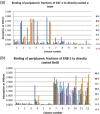
Binding of the periplasmic fractions of the master plate EAT‐1 to α toxin (a) and binding of the periplasmic fractions of the master plate ENB‐1 to NetB (b). The binding strength is indicated by absorbance at 490 nm
Figure A2
Figure A2
Alignment of the sequences of the VHHs picked from master plate EAT‐1. Conserved residues are highlighted
Figure A3
Figure A3
Alignment of the sequences of the VHHs picked from master plate ENB‐1. Conserved residues are highlighted
Figure A4
Figure A4
Neutralization of nanobodies α toxin hemolysis by VHH antibodies. A twofold dilution series of VHH antibodies, starting from 5 µM concentration, was preincubated with a commercial α toxin, after which 1% sheep erythrocytes was added. Serum derived from calves immunized with recombinant α toxin was used as a control (control serum, same immunogen as used for llama immunization). The optical density of 100% hemolysis was obtained by diluting the sheep erythrocytes in distilled water. As a control, sheep erythrocytes were incubated with the α toxin, but without VHH antibodies or serum was used. This resulted in 100% hemolysis. The data are representative of three independent experiments. VHH, Variable domain of the Heavy chain of Heavy chain
Figure A5
Figure A5
Putative ENB‐ID11_R56H and ENB‐IA4 interacting epitopes on NetB predicted based on in silico structural modeling. (a) Nanobody sequences; the regions of differences are highlighted in brown and red. (b) Homology models of VHH3 (blue) and VHH4 (purple). (c,d) Nanobody interacting epitopes on NetB (β sandwich, rim, and stem domains; shown in red)
Figure A6
Figure A6
Inhibitory activity of L. reuteri 3632 (a) and L. reuteri 3630 (b) against C. perfringens in an agar overlay assay. The data are representative of three independent experiments
Figure A7
Figure A7
Genomic organization of pyrE locus in L. reuteri 3630 and 3632 genomes. Note that both L. reuteri 3630 and 3632 have identical genomic organization; the genomic organization of only L. reuteri 3632 is shown here
Figure C1
Figure C1
Colonization of engineered L. reuteri candidates. L. reuteri strains were administered via in ovo to 18‐day embryonated chicken eggs and the birds were killed 7 days after hatching and CFUs were quantified from cecal contents. The strains were marked with rifampicin resistance to selectively isolate NE01, NE06, L. reuteri 3630, and L. reuteri 3632 from the rest of the microbiota from cecal contents. For each group, the data represent the mean ± SD of the results of five chicks

References

    1. Abildgaard, L. , Engberg, R. M. , Pedersen, K. , Schramm, A. , & Hojberg, O. (2009). Sequence variation in the alpha‐toxin encoding plc gene of Clostridium perfringens strains isolated from diseased and healthy chickens. Veterinary Microbiology, 136, 293–299. - PubMed
    1. Alimolaei, M. , Golchin, M. , & Ezatkhah, M. (2017). Orally administered recombinant Lactobacillus casei vector vaccine expressing beta‐toxoid of Clostridium perfringens that induced protective immunity responses. Research in Veterinary Science, 115, 332–339. - PubMed
    1. Allain, T. , Mansour, N. M. , Bahr, M. M. , Martin, R. , Florent, I. , Langella, P. , & Bermudez‐Humaran, L. G. (2016). A new lactobacilli in vivo expression system for the production and delivery of heterologous proteins at mucosal surfaces. FEMS Microbiology Letters, 363(13), fnw117. - PubMed
    1. Al‐Sheikhly, F. , & Truscott, R. B. (1977a). The pathology of necrotic enteritis of chickens following infusion of broth cultures of Clostridium perfringens into the duodenum. Avian Diseases, 21, 230–240. - PubMed
    1. Al‐Sheikhly, F. , & Truscott, R. B. (1977b). The pathology of necrotic enteritis of chickens following infusion of crude toxins of Clostridium perfringens into the duodenum. Avian Diseases, 21, 241–255. - PubMed

MeSH terms

LinkOut - more resources