Review
. 2022 Apr 11:10:857066.
doi: 10.3389/fcell.2022.857066. eCollection 2022.

Clinical Potential of Dental Pulp Stem Cells in Pulp Regeneration: Current Endodontic Progress and Future Perspectives

Affiliations
Review

Clinical Potential of Dental Pulp Stem Cells in Pulp Regeneration: Current Endodontic Progress and Future Perspectives

Kyu Hwan Kwack et al. Front Cell Dev Biol. .

Abstract

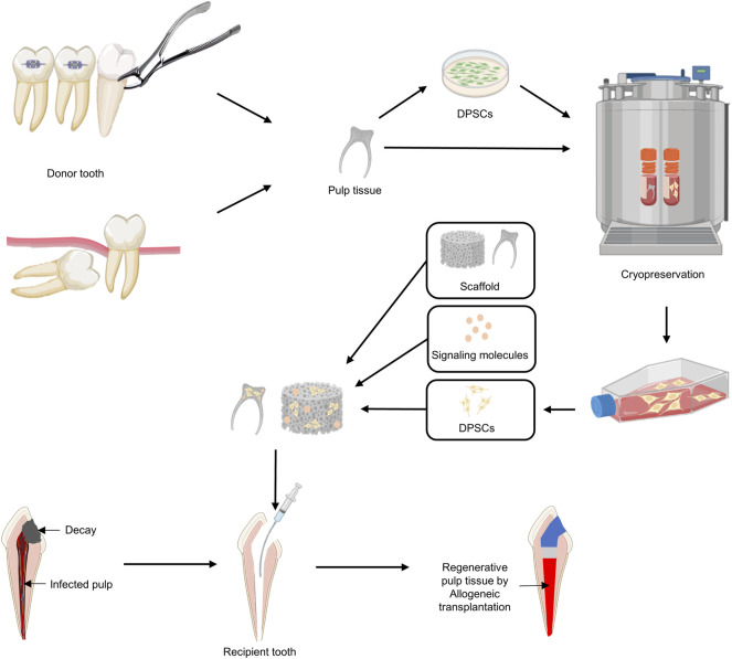
Dental caries is a common disease that not only destroys the rigid structure of the teeth but also causes pulp necrosis in severe cases. Once pulp necrosis has occurred, the most common treatment is to remove the damaged pulp tissue, leading to a loss of tooth vitality and increased tooth fragility. Dental pulp stem cells (DPSCs) isolated from pulp tissue exhibit mesenchymal stem cell-like characteristics and are considered ideal candidates for regenerating damaged dental pulp tissue owing to their multipotency, high proliferation rate, and viability after cryopreservation. Importantly, DPSCs do not elicit an allogeneic immune response because they are non-immunogenic and exhibit potent immunosuppressive properties. Here, we provide an up-to-date review of the clinical applicability and potential of DPSCs, as well as emerging trends in the regeneration of damaged pulp tissue. In addition, we suggest the possibility of using DPSCs as a resource for allogeneic transplantation and provide a perspective for their clinical application in pulp regeneration.

Keywords: allogeneic transplantation; dental pulp regeneration; dental pulp stem cells; immunomodulation; mesenchymal stem cells; regeneration medicine.


Conflict of interest statement

The authors declare that the research was conducted in the absence of any commercial or financial relationships that could be construed as a potential conflict of interest.

Figures

FIGURE 1
FIGURE 1
Schematic diagram of the current concept of pulp therapy. (A) Direct pulp capping. Pulp capping is a method used to prevent necrosis of the dental pulp when it is slightly exposed to the pin-point. Direct pulp capping covers the exposed dental pulp with a base that protects the pulp and prevents infection to maintain dental pulp vitality. (B) Pulpotomy. Pulpotomy is a minimally invasive method that is clinically considered when there is pulpitis in the absence of root pathology. (C) Pulpectomy. Pulpectomy refers to a root canal treatment that removes irreversibly infected or necrotic pulp tissue.
FIGURE 2
FIGURE 2
Characteristics of dental pulp stem cells (DPSCs). DPSCs can be isolated from dental pulp tissue and express markers similar to those on mesenchymal stem cells (MSCs). DPSCs can undergo self-renewal and have the potential to differentiate into ectoderm, mesoderm, and endoderm.
FIGURE 3
FIGURE 3
Schematic diagram of the immunosuppressive potential of dental pulp stem cells (DPSCs). (A) DPSCs can inhibit the proliferation of natural killer (NK), T helper 1 (Th1), and T helper 2 (Th2) cells and the secretion of pro-inflammatory factors such as TNF-α. In addition, DPSCs induce the proliferation of regulatory T cells (Tregs)/T helper 17 (Th17) cells and differentiation toward macrophage M2 phenotype and secrete anti-inflammatory factors such as TGF-β. (B) Fas ligand expressed in DPSCs induces T-cell apoptosis through Fas apoptotic pathway. IFN-γ secreted from hyper-activated T cells primes DPSCs to secrete TGF-β, resulting in immunosuppressive ability.
FIGURE 4
FIGURE 4
Schematic diagram of current approaches for pulp tissue regeneration. (A) Classical endodontic treatment. (B) Pulp revascularization. (C) Cell-homing-based regenerative endodontic treatment (RET). (D) Cell-transplantation-based RET.
FIGURE 5
FIGURE 5
A proposed strategy using allogeneic DPSC transplantation for pulp regeneration. 1) Dental pulp tissue is removed from an uninfected orthodontic extraction tooth or wisdom tooth (donor). 2) After isolating and culturing DPSCs from dental pulp, cryopreservation of DPSCs or dental pulp itself is performed. 3) DPSCs are expanded using cryopreserved DPSCs or dental pulp tissues according to the patient’s treatment plan for pulpectomy. 4) Signaling molecules and DPSCs are added to the appropriate scaffold and applied to the disinfected pulp space.

References

    1. Abbass M. M. S., El-Rashidy A. A., Sadek K. M., Moshy S. E., Radwan I. A., Rady D., et al. (2020). Hydrogels and Dentin-Pulp Complex Regeneration: From the Benchtop to Clinical Translation. Polymers 12 (12), 2935. 10.3390/polym12122935 - DOI - PMC - PubMed
    1. Ahmed G. M., Abouauf E. A., AbuBakr N., Dörfer C. E., El-Sayed K. F. (2020). Tissue Engineering Approaches for Enamel, Dentin, and Pulp Regeneration: An Update. Stem Cell Int. 2020, 1–15. 10.1155/2020/5734539 - DOI - PMC - PubMed
    1. Ankrum J. A., Ong J. F., Karp J. M. (2014). Mesenchymal Stem Cells: Immune Evasive, Not Immune Privileged. Nat. Biotechnol. 32 (3), 252–260. 10.1038/nbt.2816 - DOI - PMC - PubMed
    1. Apel C., Forlenza O. V., de Paula V. J. R., Talib L. L., Denecke B., Eduardo C. P., et al. (2009). The Neuroprotective Effect of Dental Pulp Cells in Models of Alzheimer’s and Parkinson’s Disease. J. Neural Transm. 116 (1), 71–78. 10.1007/s00702-008-0135-3 - DOI - PubMed
    1. Arthur A., Rychkov G., Shi S., Koblar S. A., Gronthos S. (2008). Adult Human Dental Pulp Stem Cells Differentiate toward Functionally Active Neurons Under Appropriate Environmental Cues. Stem Cells 26 (7), 1787–1795. 10.1634/stemcells.2007-0979 - DOI - PubMed

LinkOut - more resources