Review
. 2021 Jul 9;11(39):24254-24281.
doi: 10.1039/d1ra03289e. eCollection 2021 Jul 6.

Preparation and application of layered double hydroxide nanosheets

Affiliations
Review

Preparation and application of layered double hydroxide nanosheets

Yaping Zhang et al. RSC Adv. .

Abstract

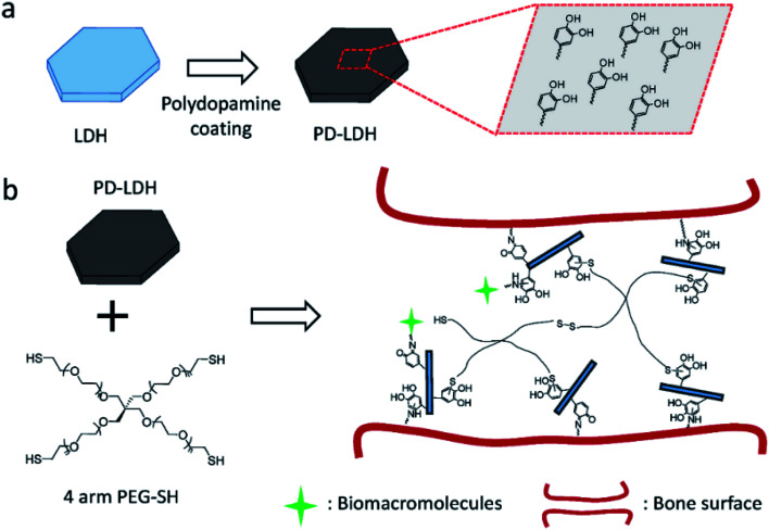
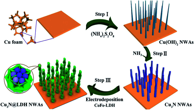
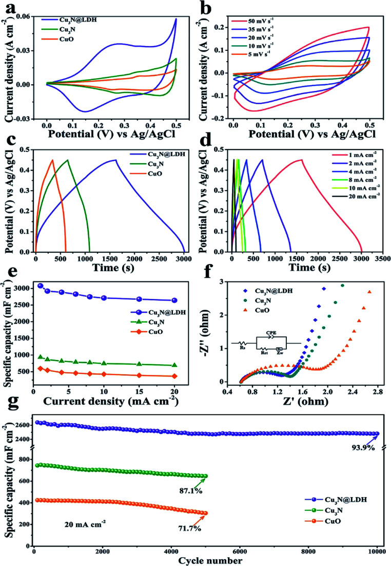
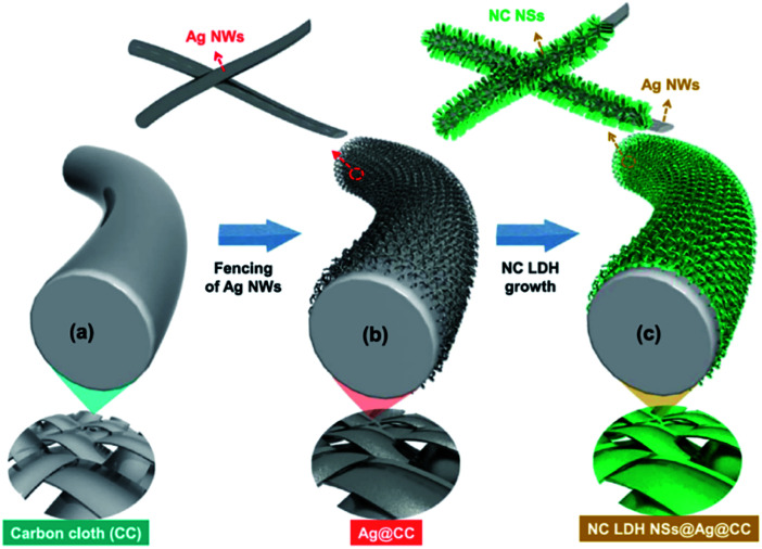
Layered double hydroxides (LDH) with unique structure and excellent properties have been widely studied in recent years. LDH have found widespread applications in catalysts, polymer/LDH nanocomposites, anion exchange materials, supercapacitors, and fire retardants. The exfoliated LDH ultrathin nanosheets with a thickness of a few atomic layers enable a series of new opportunities in both fundamental research and applications. In this review, we mainly summarize the LDH exfoliation methods developed in recent years, the recent developments for the direct synthesis of LDH single-layer nanosheets, and the applications of LDH nanosheets in catalyzing oxygen evolution reactions, crosslinkers, supercapacitors and delivery carriers.


Conflict of interest statement

The authors declare that they have no known competing financial interests or personal relationships that could have appeared to influence the work reported in this paper.

Figures

Fig. 1
Fig. 1. A general process of exfoliating LDH.
Fig. 2
Fig. 2. (A) TEM image, (B) HRTEM image and (C) SAED pattern of the exfoliated Co–Al LDH nanosheets. Reproduced with permission from ref. . Copyright 2013. The Royal Society of Chemistry.
Fig. 3
Fig. 3. The AFM image, 3D surface image and height profile of exfoliated LDH. (a) MgAl-SC-LDH; (b) MgAl-SL-LDH. Reproduced with permission from ref. . Copyright 2019. Taylor & Francis Group, LLC.
Fig. 4
Fig. 4. The schematic diagram of delamination of LDH in precooled NaOH/urea aqueous solution. Reproduced with permission from ref. . Copyright 2014 The Royal Society of Chemistry.
Fig. 5
Fig. 5. SEM, TEM and HRTEM images of the bulk CoFe LDH (A–C) and the ultrathin CoFe LDH-Ar nanosheets (D–F). Reproduced with permission from ref. . Copyright 2017 John Wiley & Sons, Inc.
Fig. 6
Fig. 6. AFM images of (A) bulk CoFe LDH and (B) ultrathin CoFe LDH-Ar nanosheets. (C) The corresponding height curves and (D) XRD patterns of the bulk CoFe LDH and ultrathin CoFe LDH-Ar nanosheets. Reproduced with permission from ref. . Copyright 2017 John Wiley & Sons, Inc.
Fig. 7
Fig. 7. Schematic representation of the nucleation and growth of LDH platelets. Reproduced with permission from ref. . Copyright 2006 The Royal Society of Chemistry.
Fig. 8
Fig. 8. (a) AFM image of the particles synthesized by a reverse micro-emulsion method deposited on a highly oriented pyrolytic graphite (HOPG) surface; (b) cross-sectional analysis of the labeled particles in (a) showing the dimensional profiles. Reproduced with permission from ref. . Copyright 2006 The Royal Society of Chemistry.
Fig. 9
Fig. 9. (A) Proposed scheme for the preparation of exfoliated Mg2/Al–LDH nanosheets; (B) AFM image of the synthesized ultrathin sheets. Reproduced with permission from ref. . Copyright 2012 Elsevier Inc.
Fig. 10
Fig. 10. (a) Comparison of the XRD patterns of Zn2Al-borate LDH synthesized by conventional co-precipitation (washing with water) and the AMOST method; (b) Mg3Al-borate LDH synthesized by conventional co-precipitation (washing with water) and the AMOST method; (c) schematic illustration of the structure of [B4O5(OH)4]2− with the LDH layers. Reflections from the sample holder. Reproduced with permission from ref. . The Royal Society of Chemistry.
Fig. 11
Fig. 11. (a) Direct growth of LDH single-layer nanosheets with the assistance of layer growth inhibitors (not drawn to scale); (b) AFM image of a pseudohexagonal Mg4/Al–LDH nanosheet. Reproduced with permission from ref. . Copyright 2015 The Royal Society of Chemistry.
Fig. 12
Fig. 12. (A) LDH characteristic peak to internal reference peak area ratios. (B) MgAl–LDH layer charge at different Mg/Al formulation molar ratios and formamide concentrations. (C) Representative AFM image of Mg2Al–LDH prepared in 30.0 vol% formamide and the corresponding height profile (D). Reproduced with permission from ref. . Copyright 2016 The American Chemistry Society.
Fig. 13
Fig. 13. Schematic illustration for formation and structure of CoAl–CO32− SL-LDH and CoAl–CO32− LDH: (a) ethylene glycol (EG) solution of reactants; (b) adsorption of EG molecules on the surface of a SL-LDH nanosheet in reaction; (c) completely dispersed SL-LDH in water; (d) disorderly stack of SL-LDH in the dried state; and (e) LDH assembly structure from generated SL-LDH. Reproduced with permission from ref. . Copyright 2017 The American Chemistry Society.
Fig. 14
Fig. 14. (a) XRD patterns of different samples and (b) AFM image of LDH-H2O. Reproduced with permission from ref. . Copyright 2017 The American Chemistry Society.
Fig. 15
Fig. 15. TEM images of Mg1.97/Fe and Co0.96/Fe–LDH materials. Scale bars in the Mg1.97/Fe–LDH images, (a and b), represent 200 nm and 50 nm, respectively. Scale bars in the Co0.96/Fe–LDH images, (c and d), represent 1 mm and 50 nm, respectively. (e) Co1.07/Al and (f) Zn1.48/Al–LDH nanosheets. The dotted arrows marked by symbol “A” indicate the edges of the nanosheets seriously rolled because of the instability of the edges of the free-standing ultrathin LDH layers. Both scale bars represent 50 nm in (e and f). Insets in images (b, d, and f) display the corresponding side views. Reproduced with permission from ref. . Copyright 2010 American Institute of Physics.
Fig. 16
Fig. 16. (A) AFM image and height scale of the LDH nanosheets, and (B) sectional analysis along the black line marked in (A). Reproduced with permission from ref. . Copyright 2014 Elsevier Inc.
Fig. 17
Fig. 17. TEM images of dispersions of (A) LDH single layer nanosheets gel, (B) LDH single layer nanosheets, (E) LDH single layer nanosheets after stacking at ∼25 °C, and (F) peptized LDH platelets; (C) HR-TEM image and (D) SAED pattern of LDH single layer nanosheets; (G) AFM image and sectional analysis of the LDH single layer nanosheets. Reproduced with permission from ref. . Copyright 2016 Elsevier Inc.
Fig. 18
Fig. 18. AFM image (a) and height profile (b) of Co–Ni LDH monolayer nanosheets prepared at molar ratio of Co2+ to Ni2+ = 0.2. Reproduced with permission from ref. . Copyright 2016 Elsevier Inc.
Fig. 19
Fig. 19. Schematic illustration for the structure of multilayered Co–Ni LDH (a) and the formation mechanism of Co–Ni LDH monolayer nanosheets (b). Reproduced with permission from ref. . Copyright 2016 Elsevier Inc.
Fig. 20
Fig. 20. (A) TEM, (B) HAADF-STEM, and (C) HRTEM image of PM-LDH; (D) the FFT pattern of the image shown in (C). (E) AFM image and (F) AFM height profiles of PM-LDH; the numbers 1–3 in (E) correspond to the profiles 1–3 in (F). (G) A dispersion of PM-LDH in ethanol displaying the Tyndall effect. Reproduced with permission from ref. . Copyright 2019 John Wiley & Sons, Inc.
Fig. 21
Fig. 21. (a) LSV curves for OER measured at a scan rate of 5 mV s−1; (b) the overpotential of ZnCo-UF at the current density of 5 mA cm−2; (c) corresponding Tafel plots; (d) EIS Nyquist plots at 1.8 V. Reproduced with permission from ref. . Copyright 2019 Elsevier Inc.
Fig. 22
Fig. 22. (a) LSV curves for OER on bulk NiMn-LDH and ultrathin NiMn-LDH nanosheets. (b) The corresponding Tafel plots. (c) Cycles stability of the exfoliated NiMn-LDH nanosheets. (d) DFT calculated free energy diagrams for the OER process from H2O to O2 on the NiMn-LDH structures. The optimized configurations of the adsorption intermediates of *OH, *O and *OOH are shown inside. Dark blue: Mn; green: Ni; red: O; white: H. Reproduced with permission from ref. . Copyright 2018 John Wiley & Sons, Inc.
Fig. 23
Fig. 23. The OER performance of bulk CoFe LDH and ultrathin CoFe LDH-Ar nanosheets. (A) LSV curves for the OER at a scan rate of 5 mV s−1. (B) The corresponding Tafel plots. (C) Nyquist plots at an overpotential of 270 mV. The inset gives the equivalent circuit. Rs = series resistance, Rp = charge transfer resistance, CPE = constant phase element related to the double-layer capacitance. (D) Stability test with the CoFe LDH-Ar on Ni foam. Reproduced with permission from ref. . Copyright 2017 John Wiley & Sons, Inc.
Fig. 24
Fig. 24. Typical SEM images of the freeze-dried xerogels for the neat PAM hydrogel (a) and the LDH/PAM NC hydrogel (L1M) (b–d). The neat PAM xerogel showed the common large-pore structure at the micrometer scale (a). In contrast, the NC xerogel revealed an unusual hierarchical porous structure with interconnected pore sizes at micro- and nanometer scales (b and c). Inset in (c): enlargement of the outlined rectangular part. (d) Details of the nanometer-sized pores. Reproduced with permission from ref. . Copyright 2014 John Wiley & Sons, Inc.
Fig. 25
Fig. 25. Stress–strain curves of (a) tensile and (b) compression measurements for the as-prepared LDH/PAM NC hydrogels (LmM). Reproduced with permission from ref. . Copyright 2015 The Royal Society of Chemistry.
Fig. 26
Fig. 26. (a) Schematic representation of polydopamine coating on layered double hydroxides (LDH). (b) Schematic of nanocomposite hydrogel synthesized with 4 arm thiol-terminated polyethylene glycol (4 arm PEG-SH) and polydopamine-coated LDH (PD-LDH), which is able to immobilize biomacromolecules and act as the bioadhesive for bone fracture. Reproduced with permission from ref. . Copyright 2016 John Wiley & Sons, Inc.
Fig. 27
Fig. 27. Schematic illustration for the fabrication of hierarchical Cu3N@CoFe-LDH core–shell NWAs supported on copper foam. Reproduced with permission from ref. . Copyright 2018 The Royal Society of Chemistry.
Fig. 28
Fig. 28. (a) CV curves of CuO, Cu3N and Cu3N@CoFe-LDH at a scan rate of 5 mV s−1. (b) CV curves of Cu3N@CoFe-LDH at various scan rates. (c) GCD curves of CuO, Cu3N and Cu3N@CoFe-LDH at a current density of 1 mA cm−2. (d) GCD curves of Cu3N@CoFe-LDH at various current densities. (e) Current density dependence of the areal capacitance, (f) Nyquist plots of EIS and (g) cycling performance of the pristine CuO, Cu3N and Cu3N@CoFe-LDH. Reproduced with permission from ref. . Copyright 2018 The Royal Society of Chemistry.
Fig. 29
Fig. 29. (a–c) Schematic illustration showing the preparation process flow of NC LDH NSs@Ag@CC by a single-step ECD process. Reproduced with permission from ref. . Copyright 2017 Elsevier Inc.
Fig. 30
Fig. 30. Characterizations for the monolayer-NiTi-LDH nanosheets. (A) TEM image; (B) HRTEM image; (C) AFM image and (D) the corresponding height profiles; the numbers from 1 to 3 in (D) correspond to the numbers from 1 to 3 in (C). Reproduced with permission from ref. . Copyright 2015 The Royal Society of Chemistry.
Fig. 31
Fig. 31. (a) XRD patterns of bulk LDH colloid (black line), MLDH colloid (red line), and the re-stacking of MLDH nanosheets (blue line). (b) AFM image, (c) HRTEM image, and (d) particle size distribution of MLDH nanosheets. The insets in (c) show a high magnification image and crystal lattice. Reproduced with permission from ref. . Copyright 2018 Springer.
Fig. 32
Fig. 32. (a) A schematic illustration for MLDH-based drug delivery system toward efficient loading and precisely controlled delivery of theranostic agents. (b) HRTEM image of MLDH nanosheets. (c) AFM image and (d) measured thickness of MLDH nanosheets. (e) Size distribution of MLDH nanosheets in water, PBS, and culture medium (high glucose Dulbecco's modified Eagle medium (DMEM)). (f) XRD patterns of: (1) bulk LDH colloid, (2) MLDH nanosheets colloid, and (3) the restacking sample of MLDH nanosheets. The peak at 26.2° is ascribed to the PET film substrate. (g) XPS spectra of MLDH nanosheets sample. Reproduced with permission from ref. . Copyright 2018 John Wiley & Sons, Inc.
Fig. 33
Fig. 33. In vivo anticancer activities: (a) tumor growth curves of 4T1 tumor-bearing BALB/C mice after intravenous injection with (1) PBS, (2) free MTX (MTX), (3) MTX loaded in traditional LDH nanoparticles (LDH-MTX), and (4) MTX loaded in PEG-modified ultrathin LDH nanosheets (LDH-Co-PEG MTX) (n = 5, error bar indicates standard deviation; the arrow indicates tail vein injection at day 1; * indicates p < 0.05 with Student's t test). (b) The body weight evolution of 4T1 tumor-bearing mice at different times after intravenous injection of different materials (n = 5, error bar indicates standard deviation). (c) H&E staining images of tumors slices. The tumor-bearing mice were treated with MTX, LDH-MTX, and LDH-Co-PEG MTX at day 1 and sacrificed at day 7. Higher cancer cell damage (red area indicates area of cell damage) was found in LDH MTX- and LDH-Co-PEG MTX-treated groups. Scale bar: 100 μm. Reproduced with permission from ref. . Copyright 2017 The American Chemistry Society.

References

    1. Sideris P. J. Nielsen U. G. Gan Z. H. Grey C. P. Science. 2008;321:113–117. doi: 10.1126/science.1157581. - DOI - PubMed
    1. Ma R. Z. Liu Z. P. Takada K. Iyi N. Bando Y. Sasaki T. J. Am. Chem. Soc. 2007;129:5257–5263. doi: 10.1021/ja0693035. - DOI - PubMed
    1. Wang Q. O'Hare D. Chem. Rev. 2012;112:4124–4155. doi: 10.1021/cr200434v. - DOI - PubMed
    1. Liu Z. P. Ma R. Z. Ebina Y. Iyi N. Takada K. Sasaki T. Langmuir. 2007;23:861–867. doi: 10.1021/la062345m. - DOI - PubMed
    1. Gomes Silva C. Bouizi Y. Fornes V. Garcia H. J. Am. Chem. Soc. 2009;131:13833. doi: 10.1021/ja905467v. - DOI - PubMed