

Metal Nanoparticle Modified Carbon-Fiber Microelectrodes Enhance Adenosine Triphosphate Surface Interactions with Fast-Scan Cyclic Voltammetry
- PMID: 35479102
- PMCID: PMC9026253
- DOI: 10.1021/acsmeasuresciau.1c00026
Metal Nanoparticle Modified Carbon-Fiber Microelectrodes Enhance Adenosine Triphosphate Surface Interactions with Fast-Scan Cyclic Voltammetry
Abstract
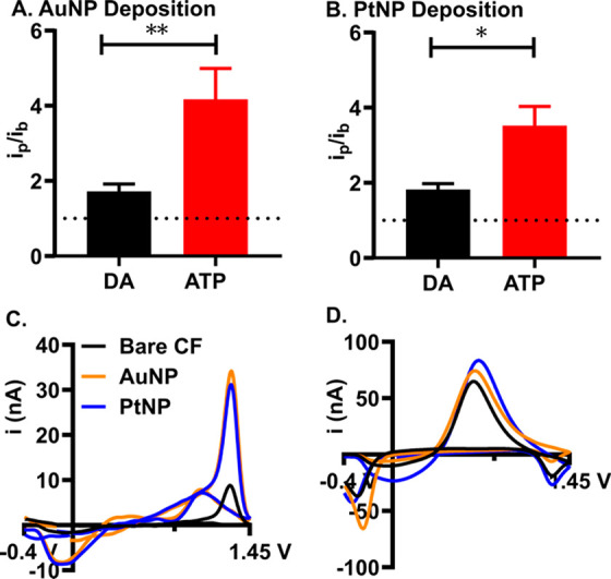
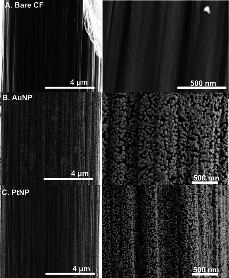
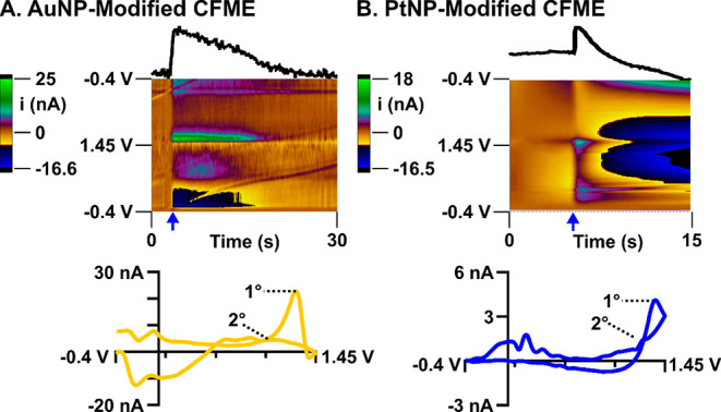
Adenosine triphosphate (ATP) is an important rapid signaling molecule involved in a host of pathologies in the body. Historically, ATP is difficult to directly detect electrochemically with fast-scan cyclic voltammetry (FSCV) due to limited interactions at bare carbon-fibers. Systematic investigations of how ATP interacts at electrode surfaces is necessary for developing more sensitive electrochemical detection methods. Here, we have developed gold nanoparticle (AuNP), and platinum nanoparticle (PtNP) modified carbon-fiber microelectrodes coupled to FSCV to measure the extent to which ATP interacts at metal nanoparticle-modified surfaces and to improve the sensitivity of direct electrochemical detection. AuNP and PtNPs were electrodeposited on the carbon-fiber surface by scanning from -1.2 to 1.5 V for 30 s in 0.5 mg/mL HAuCl4 or 0.5 mg/mLK2PtCl6. Overall, we demonstrate an average 4.1 ± 1.0-fold increase in oxidative ATP current at AuNP-modified and a 3.5 ± 0.3-fold increase at PtNP-modified electrodes. Metal nanoparticle-modified surfaces promoted improved electrocatalytic conversion of ATP oxidation products at the surface, facilitated enhanced adsorption strength and surface coverage, and significantly improved sensitivity. ATP was successfully detected within living murine lymph node tissue following exogenous application. Overall, this study demonstrates a detailed characterization of ATP oxidation at metal nanoparticle surfaces and a significantly improved method for direct electrochemical detection of ATP in tissue.
© 2021 The Authors. Published by American Chemical Society.
Conflict of interest statement
The authors declare no competing financial interest.
Figures


Similar articles
-
Platinum Nanoparticle Size and Density Impacts Purine Electrochemistry with Fast-Scan Cyclic Voltammetry.J Electrochem Soc. 2022 Apr;169(4):046514. doi: 10.1149/1945-7111/ac65bc. Epub 2022 Apr 19. J Electrochem Soc. 2022. PMID: 35497383 Free PMC article.
-
Amine-functionalized carbon-fiber microelectrodes for enhanced ATP detection with fast-scan cyclic voltammetry.Anal Methods. 2021 May 27;13(20):2320-2330. doi: 10.1039/d1ay00089f. Anal Methods. 2021. PMID: 33960336 Free PMC article.
-
Gold Nanoparticle Modified Carbon Fiber Microelectrodes for Enhanced Neurochemical Detection.J Vis Exp. 2019 May 13;(147):10.3791/59552. doi: 10.3791/59552. J Vis Exp. 2019. PMID: 31132067 Free PMC article.
-
Fast-scan Cyclic Voltammetry for the Characterization of Rapid Adenosine Release.Comput Struct Biotechnol J. 2014 Dec 29;13:47-54. doi: 10.1016/j.csbj.2014.12.006. eCollection 2015. Comput Struct Biotechnol J. 2014. PMID: 26900429 Free PMC article. Review.
-
Recent advances in fast-scan cyclic voltammetry.Analyst. 2020 Feb 17;145(4):1087-1102. doi: 10.1039/c9an01925a. Analyst. 2020. PMID: 31922162 Free PMC article. Review.
Cited by
-
State of the art indicators for imaging purinergic dynamics in vitro and in vivo.Purinergic Signal. 2025 Jun 17. doi: 10.1007/s11302-025-10095-6. Online ahead of print. Purinergic Signal. 2025. PMID: 40526259 Review.
-
Editors' Choice-Review-The Future of Carbon-Based Neurochemical Sensing: A Critical Perspective.ECS Sens Plus. 2023 Dec 1;2(4):043601. doi: 10.1149/2754-2726/ad15a2. Epub 2023 Dec 27. ECS Sens Plus. 2023. PMID: 38170109 Free PMC article. Review.
-
Fibrous wearable and implantable bioelectronics.Appl Phys Rev. 2023 Sep;10(3):031303. doi: 10.1063/5.0152744. Appl Phys Rev. 2023. PMID: 37576610 Free PMC article. Review.
-
Wet-Spun Porous Carbon Microfibers for Enhanced Electrochemical Detection.ACS Appl Mater Interfaces. 2023 Apr 12;15(14):17601-17611. doi: 10.1021/acsami.3c00423. Epub 2023 Mar 29. ACS Appl Mater Interfaces. 2023. PMID: 36989172 Free PMC article.
-
Potentiometric MIP-Modified Screen-Printed Cell for Phenoxy Herbicides Detection.Int J Environ Res Public Health. 2022 Dec 8;19(24):16488. doi: 10.3390/ijerph192416488. Int J Environ Res Public Health. 2022. PMID: 36554364 Free PMC article.
References
-
- Gardner A.; Johansson A.; Wibom R.; Nennesmo I.; von Döbeln U.; Hagenfeldt L.; Hällström T. Alterations of Mitochondrial Function and Correlations with Personality Traits in Selected Major Depressive Disorder Patients. J. Affective Disord. 2003, 76 (1), 55–68. 10.1016/S0165-0327(02)00067-8. - DOI - PubMed
Grants and funding
LinkOut - more resources
Full Text Sources