. 2022 Apr 5:13:804168.
doi: 10.3389/fpsyt.2022.804168. eCollection 2022.

Sex Modulates the Pathological Aging Effect on Caudate Functional Connectivity in Mild Cognitive Impairment

Collaborators, Affiliations

Sex Modulates the Pathological Aging Effect on Caudate Functional Connectivity in Mild Cognitive Impairment

Zhengshi Yang et al. Front Psychiatry. .

Abstract

Purpose: To assess the pathological aging effect on caudate functional connectivity among mild cognitive impairment (MCI) participants and examine whether and how sex and amyloid contribute to this process.

Materials and methods: Two hundred and seventy-seven functional magnetic resonance imaging (fMRI) sessions from 163 cognitive normal (CN) older adults and 309 sessions from 139 participants with MCI were included as the main sample in our analysis. Pearson's correlation was used to characterize the functional connectivity (FC) between caudate nuclei and each brain region, then caudate nodal strength was computed to quantify the overall caudate FC strength. Association analysis between caudate nodal strength and age was carried out in MCI and CN separately using linear mixed effect (LME) model with covariates (education, handedness, sex, Apolipoprotein E4, and intra-subject effect). Analysis of covariance was conducted to investigate sex, amyloid status, and their interaction effects on aging with the fMRI data subset having amyloid status available. LME model was applied to women and men separately within MCI group to evaluate aging effects on caudate nodal strength and each region's connectivity with caudate nuclei. We then evaluated the roles of sex and amyloid status in the associations of neuropsychological scores with age or caudate nodal strength. An independent cohort was used to validate the sex-dependent aging effects in MCI.

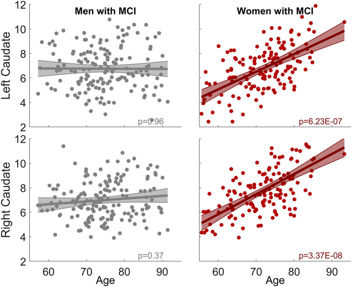
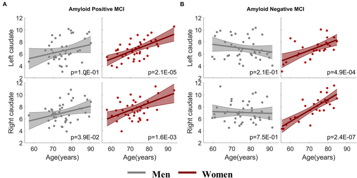
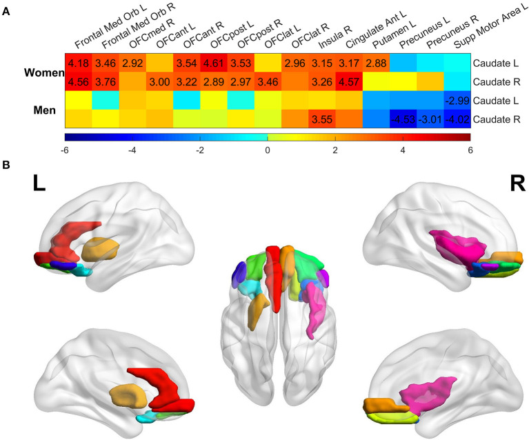
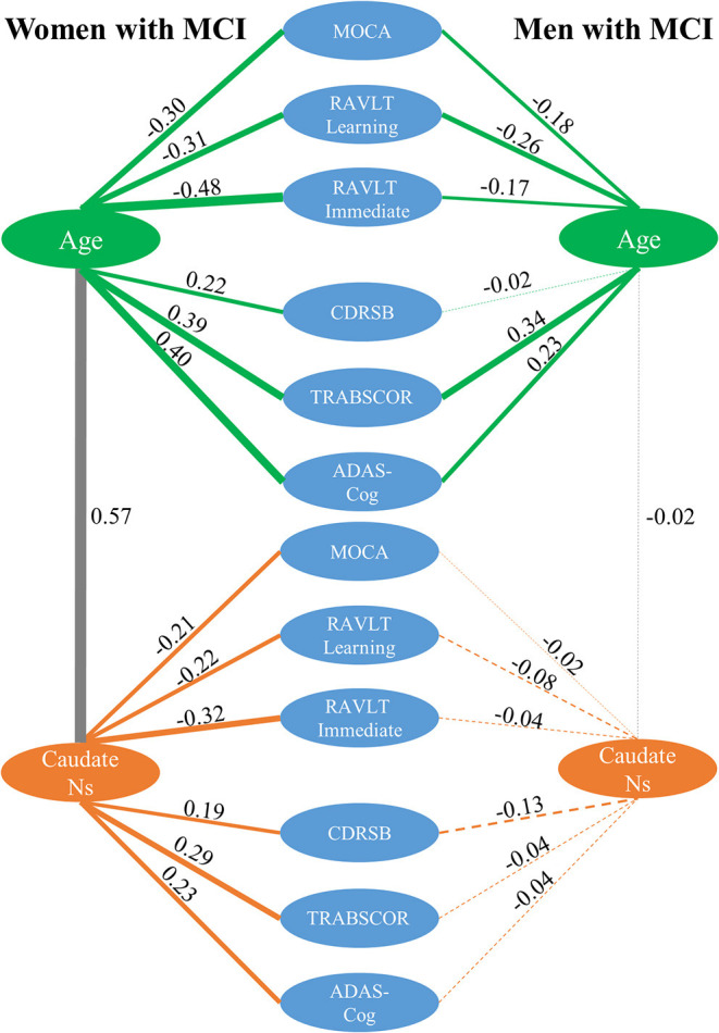
Results: The MCI group had significantly stronger age-related increase of caudate nodal strength compared to the CN group. Analyzing women and men separately revealed that the aging effect on caudate nodal strength among MCI participants was significant only for women (left: P = 6.23 × 10⁻⁷, right: P = 3.37 × 10⁻⁸), but not for men (P > 0.3 for bilateral caudate nuclei). The aging effects on caudate nodal strength were not significantly mediated by brain amyloid burden. Caudate connectivity with ventral prefrontal cortex substantially contributed to the aging effect on caudate nodal strength in women with MCI. Higher caudate nodal strength is significantly related to worse cognitive performance in women but not in men with MCI.

Conclusion: Sex modulates the pathological aging effects on caudate nodal strength in MCI regardless of amyloid status. Caudate nodal strength may be a sensitive biomarker of pathological aging in women with MCI.

Keywords: Alzheimer's Disease; aging effect; caudate; functional connectivity; mild cognitive impairment; sex difference.


Conflict of interest statement

The authors declare that the research was conducted in the absence of any commercial or financial relationships that could be construed as a potential conflict of interest.

Figures

Figure 1
Figure 1
Associations between bilateral caudate nodal strength and age. (A) Priori-defined bilateral caudate used in the analysis. (B) Scatter plot of the nodal strength from left/right caudate with age for MCI and CN separately. Shaded area represents 95% confidence interval of the fitting curve. The nodal strength shown in the figure has been adjusted to remove the influence of confounding factors using LME model; see Methods section for detail. Stronger age-related increase of caudate nodal strength is observed in MCI compared to CN.
Figure 2
Figure 2
Sex-dependent association between caudate nodal strength and age among MCI group. No association is observed in men for both right and left caudate. In contrast, significant positive association is observed in women (left: p = 6.23 × 10⁻⁷, right: p = 3.37 × 10⁻⁸). The same analysis among CN group is shown in Supplementary Figure 1.
Figure 3
Figure 3
Sex-dependent association analysis between caudate nodal strength and age with stratified amyloid status. (A) Association analysis within amyloid positive MCI participants. (B) Association analysis within amyloid negative MCI participants. The x-axis is age and the y-axis is the corrected caudate nodal strength after adjusting the influence of confounding factors.
Figure 4
Figure 4
Regional analysis of aging effect on functional connectivity of individual ROI with left/right caudate among the MCI group. (A) t-statistical values of aging effect on functional connectivity of individual region with left/right caudate. All regions were assessed, but only regions having association over the significance threshold p = 0.005 in at least one scenario were listed in the figure. Only the t-values with significance level p < 0.005 were marked in the plot. (B) Brain regions having significant association with age thresholded at p < 0.005 in women with MCI, regardless of the connectivity with left or right caudate.
Figure 5
Figure 5
Correlation analysis among caudate nodal strength (Ns), neuropsychological scores, and age for women and men with MCI separately. For simplicity, only left caudate nodal strength was presented in the figure. Green, gray, and orange lines represent the correlation between age and neuropsychological scores, between age and caudate nodal strength, and between caudate nodal strength and neuropsychological scores, respectively. Pearson's correlations are marked in the figure with line thickness proportional to the correlation strength. A solid line means the correlation is significant (p < 0.05) and dashed line means the correlation is not significant (p > 0.05).

References

    1. Lambon Ralph MA, Patterson K, Graham N, Dawson K, Hodges JR. Homogeneity and heterogeneity in mild cognitive impairment and Alzheimer's disease: a cross-sectional and longitudinal study of 55 cases. Brain. (2003) 126:2350–62. 10.1093/brain/awg236 - DOI - PubMed
    1. Buckner RL. Memory and executive function in aging and AD: multiple factors that cause decline and reserve factors that compensate. Neuron. (2004) 44:195–208. 10.1016/j.neuron.2004.09.006 - DOI - PubMed
    1. Hedden T, Gabrieli JD. Insights into the ageing mind: a view from cognitive neuroscience. Nat Rev Neurosci. (2004) 5:87–96. 10.1038/nrn1323 - DOI - PubMed
    1. Fjell AM, Sneve MH, Storsve AB, Grydeland H, Yendiki A, Walhovd KB. Brain events underlying episodic memory changes in aging: a longitudinal investigation of structural and functional connectivity. Cereb Cortex. (2016) 26:1272–86. 10.1093/cercor/bhv102 - DOI - PMC - PubMed
    1. Rieckmann A, Johnson KA, Sperling RA, Buckner RL, Hedden T. Dedifferentiation of caudate functional connectivity and striatal dopamine transporter density predict memory change in normal aging. Proc Nat Acad Sci USA. (2018) 115:10160–5. 10.1073/pnas.1804641115 - DOI - PMC - PubMed

LinkOut - more resources