

A biocompatible polypyrrole membrane for biomedical applications
- PMID: 35479716
- PMCID: PMC9031619
- DOI: 10.1039/d1ra01338f
A biocompatible polypyrrole membrane for biomedical applications
Abstract
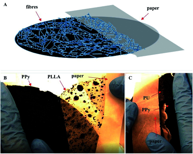
Polypyrrole (PPy) is the most widely investigated electrically conductive biomaterial. However, because of its intrinsic rigidity, PPy has only been used either in the form of a composite or a thin coating. This work presents a pure and soft PPy membrane that is synergically reinforced with the electrospun polyurethane (PU) and poly-l-lactic acid (PLLA) fibers. This particular reinforcement not only renders the originally rather fragile PPy membrane easy to manipulate, it also prevents the membrane from deformation in an aqueous environment. Peel and mechanical tests confirmed the strong adhesion of the fibers and the significantly increased tensile strength of the reinforced membrane. Surface electrical conductivity and long-term electrical stability were tested, showing that these properties were not affected by the reinforcement. Surface morphology and chemistry were analyzed with scanning electron spectroscopy (SEM), X-ray photoelectron spectroscopy (XPS), and Fourier transform infrared spectroscopy (FTIR). Material thermal stability was investigated with thermogravimetric analysis (TGA). Finally, the adhesion and proliferation of human skin keratinocytes on the membrane were assessed by Hoechst staining and the methylthiazolyldiphenyl-tetrazolium bromide (MTT) assay. In conclusion, this membrane proves to be the first PPy-based soft conductive biomaterial that can be practically used. Its electrical conductivity and cytocompatibility promise a wide range of biomedical applications.
This journal is © The Royal Society of Chemistry.
Conflict of interest statement
There are no conflicts to declare.
Figures


References
-
- Bolto B. A. McNeill R. Weiss D. E. Aust. J. Chem. 1963;16:1090–1103. doi: 10.1071/CH9631090. - DOI
-
- Street G. Lindsey S. Nazzal A. Wynne K. Mol. Cryst. Liq. Cryst. 1985;118:137–148. doi: 10.1080/00268948508076201. - DOI
-
- Vernitskaya T. V. Efimov O. N. Russ. Chem. Rev. 1997;66:443. doi: 10.1070/RC1997v066n05ABEH000261. - DOI
-
- Yilmaz F., Conducting polymers, 2016
-
- Zhang Z., Rouabhia M. and Moulton S. E., Conductive Polymers: Electrical Interactions in Cell Biology and Medicine, CRC Press, 2017
LinkOut - more resources
Full Text Sources