

Multimodal Heartbeat and Compression Optical Coherence Elastography for Mapping Corneal Biomechanics
- PMID: 35479957
- PMCID: PMC9037093
- DOI: 10.3389/fmed.2022.833597
Multimodal Heartbeat and Compression Optical Coherence Elastography for Mapping Corneal Biomechanics
Abstract
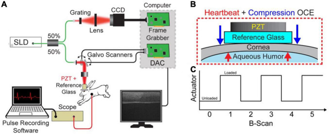
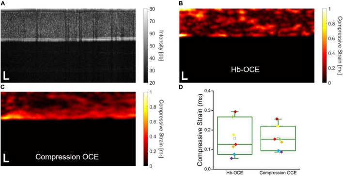
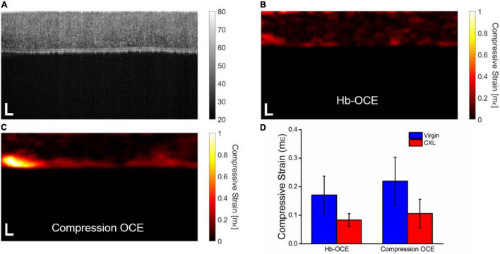
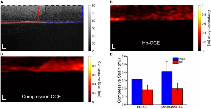
The biomechanical properties of the cornea have a profound influence on the health, structural integrity, and function of the eye. Understanding these properties may be critical for diagnosis and identifying disease pathogenesis. This work demonstrates how two different elastography techniques can be combined for a multimodal approach to measuring corneal biomechanical properties. Heartbeat optical coherence elastography (Hb-OCE) and compression OCE were performed simultaneously to measure the stiffness of the cornea in an in vivo rabbit model. Measurements were further performed after collagen crosslinking to demonstrate how the combined technique can be used to measure changes in corneal stiffness and map mechanical contrast. The results of this work further suggest that measurements from Hb-OCE and compression OCE are comparable, meaning that Hb-OCE and compression OCE may be used interchangeably despite distinct differences in both techniques.
Keywords: biomechanics; cornea; elasticity; optical coherence elastography (OCE); optical coherence tomography (OCT).
Copyright © 2022 Nair, Singh, Aglyamov and Larin.
Conflict of interest statement
The authors declare that the research was conducted in the absence of any commercial or financial relationships that could be construed as a potential conflict of interest.
Figures
Similar articles
-
Optical coherence elastography measures the biomechanical properties of the ex vivo porcine cornea after LASIK.J Biomed Opt. 2024 Jan;29(1):016002. doi: 10.1117/1.JBO.29.1.016002. Epub 2024 Jan 13. J Biomed Opt. 2024. PMID: 38223300 Free PMC article.
-
Heartbeat OCE: corneal biomechanical response to simulated heartbeat pulsation measured by optical coherence elastography.J Biomed Opt. 2020 May;25(5):1-9. doi: 10.1117/1.JBO.25.5.055001. J Biomed Opt. 2020. PMID: 32372574 Free PMC article.
-
Heartbeat optical coherence elastography: corneal biomechanics in vivo.J Biomed Opt. 2021 Feb;26(2):020502. doi: 10.1117/1.JBO.26.2.020502. J Biomed Opt. 2021. PMID: 33624461 Free PMC article.
-
Strain and elasticity imaging in compression optical coherence elastography: The two-decade perspective and recent advances.J Biophotonics. 2021 Feb;14(2):e202000257. doi: 10.1002/jbio.202000257. Epub 2020 Nov 3. J Biophotonics. 2021. PMID: 32749033 Review.
-
Anterior segment applications of optical coherence elastography in ophthalmic and vision science: a systematic review of intrinsic measurement techniques and clinical relevance.Prog Biomed Eng (Bristol). 2025 May 15;7(3). doi: 10.1088/2516-1091/add4d9. Prog Biomed Eng (Bristol). 2025. PMID: 40328290
Cited by
-
In vivo corneal elastography: A topical review of challenges and opportunities.Comput Struct Biotechnol J. 2023 Apr 13;21:2664-2687. doi: 10.1016/j.csbj.2023.04.009. eCollection 2023. Comput Struct Biotechnol J. 2023. PMID: 37181662 Free PMC article. Review.
-
Air-pulse optical coherence elastography: how excitation angle affects mechanical wave propagation.Biomed Opt Express. 2025 Mar 11;16(4):1371-1391. doi: 10.1364/BOE.557984. eCollection 2025 Apr 1. Biomed Opt Express. 2025. PMID: 40322015 Free PMC article.
-
Optical coherence elastography measures the biomechanical properties of the ex vivo porcine cornea after LASIK.J Biomed Opt. 2024 Jan;29(1):016002. doi: 10.1117/1.JBO.29.1.016002. Epub 2024 Jan 13. J Biomed Opt. 2024. PMID: 38223300 Free PMC article.
-
Multi-modal deep learning based on multi-dimensional and multi-level temporal data can enhance the prognostic prediction for multi-drug resistant pulmonary tuberculosis patients.Sci One Health. 2022 Nov 23;1:100004. doi: 10.1016/j.soh.2022.100004. eCollection 2022 Nov. Sci One Health. 2022. PMID: 39076608 Free PMC article. Review.
-
Dual optical elastography detects -induced alterations in the biomechanical properties of skin scaffolds.J Biomed Opt. 2024 Sep;29(9):095002. doi: 10.1117/1.JBO.29.9.095002. Epub 2024 Sep 18. J Biomed Opt. 2024. PMID: 39295639 Free PMC article.
References
Grants and funding
LinkOut - more resources
Full Text Sources