. 2021 Jun 23;11(36):22214-22220.
doi: 10.1039/d1ra02511b. eCollection 2021 Jun 21.

Sensing mechanism of a new fluorescent probe for hydrogen sulfide: photoinduced electron transfer and invalidity of excited-state intramolecular proton transfer

Affiliations

Sensing mechanism of a new fluorescent probe for hydrogen sulfide: photoinduced electron transfer and invalidity of excited-state intramolecular proton transfer

Xiumin Liu et al. RSC Adv. .

Abstract

It is of great significance for biological research to develop efficient detection methods of hydrogen sulfide (H2S). When DFAN reacts with H2S, 2,4-dinitrophenyl ether group acting as an electron acceptor generates a hydroxyl-substituted 2,4-dinitrophenyl ether group, resulting in the disappearance of photoinduced electron transfer (PET), and the new formed DFAH can be observed, while being accompanied by a significant fluorescence. In the present study, the PET sensing mechanism of probe DFAN and the excited state intramolecular proton transfer (ESIPT) process of DFAH have been explored in detail based on the density functional theory (DFT) and time-dependent density functional theory (TD-DFT) methods. Our theoretical results show that the fluorescence quenching of DFAN is caused by the PET mechanism, and the result of ESIPT mechanism is not due to the large Stokes shift fluorescence emission of DFAH. We also optimized the geometric structure of the transition state of DFAH. The frontier molecular orbitals and potential barrier show that the ESIPT process does not easy occur easily for DFAH. The enol structure of DFAH is more stable than that of the keto structure. The absence of the PET process resulted in the enol structure emitting strong fluorescence, which is consistent with the single fluorescence in the experiment. Above all, our calculations are sufficient to verify the sensing mechanism of H2S using DFAN.


Conflict of interest statement

There are no conflicts to declare.

Figures

Scheme 1
Scheme 1. Sensing mechanism of H2S by probe DFAN.
Fig. 1
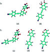
Fig. 1. Optimized geometries of DFAN and DFAH in the S0 and S1 states. (a) and (b) The S0 states of DFAN and DFAH-enol, respectively. (c)–(e) The S1 states of DFAN, DFAH-enol and DFAH-keto, respectively. The labeling of atomic color: C: blue; H: gray; O: red; N: purple.
Fig. 2
Fig. 2. Calculated IR spectra of the normal form of DFAH in the relevant spectral region of the O1–H2 stretching band in the S0 and S1 states.
Fig. 3
Fig. 3. The visual diagram of the RDG isosurface and color gradient axis. Blue indicates strong attractive interactions, and red indicates a strong bond-free overlap.
Fig. 4
Fig. 4. Potential energy curves of the S0 and S1 states of DFAH as a function of the O3–H2 bond length.
Fig. 5
Fig. 5. The calculated absorption and fluorescence spectra of DFAN and DFAH based on the TD-DFT/B3LYP/TZVP level. The corresponding experimental values are given in the parenthesis.
Fig. 6
Fig. 6. Mulliken charge distribution of molecule DFAH in S0 and S1 states. The numbers in the parentheses represent the S0 state.
Fig. 7
Fig. 7. The electron density isosurface mapped with the molecular electrostatic potential surface (MEPs) for DFAN and DFAH.
Fig. 8
Fig. 8. Calculated frontier molecular orbitals involved in the absorption of DFAN and DFAH based on B3LYP/TZVP levels (HOMO, LUMO, LUMO+1 and LUMO+2).
Fig. 9
Fig. 9. Calculated frontier molecular orbitals involved in the emission of DFAN and DFAH based on B3LYP/TZVP levels (HOMO, LUMO and LUMO+2).

References

    1. Chen L. Y. Wu D. Lim C. S. Kim D. Nam S. J. Lee W. Kim G. Kim H. M. Yoon J. Chem. Commun. 2017;53:4791–4794. doi: 10.1039/C7CC01695F. - DOI - PubMed
    1. Wallace J. L. Wang R. Nat. Rev. Drug Discovery. 2015;14:329–345. doi: 10.1038/nrd4433. - DOI - PubMed
    1. Kimura H. Nihon Yakurigaku Zasshi. 2012;139:6–8. doi: 10.1254/fpj.139.6. - DOI - PubMed
    1. Zanardo R. C. O. Brancaleone V. Distrutti E. Fiorucci S. Cirino G. Wallace J. L. FASEB J. 2006;20:2118–2120. doi: 10.1096/fj.06-6270fje. - DOI - PubMed
    1. Kimura Y. Goto Y. I. Kimura H. Antioxid. Redox Signaling. 2010;12:1–13. doi: 10.1089/ars.2008.2282. - DOI - PubMed