

Promoting osteointegration effect of Cu-alloyed titanium in ovariectomized rats
- PMID: 35480856
- PMCID: PMC9039496
- DOI: 10.1093/rb/rbac011
Promoting osteointegration effect of Cu-alloyed titanium in ovariectomized rats
Abstract
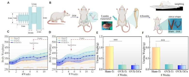
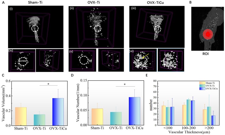
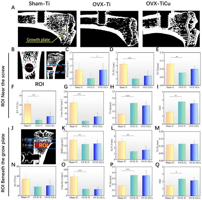
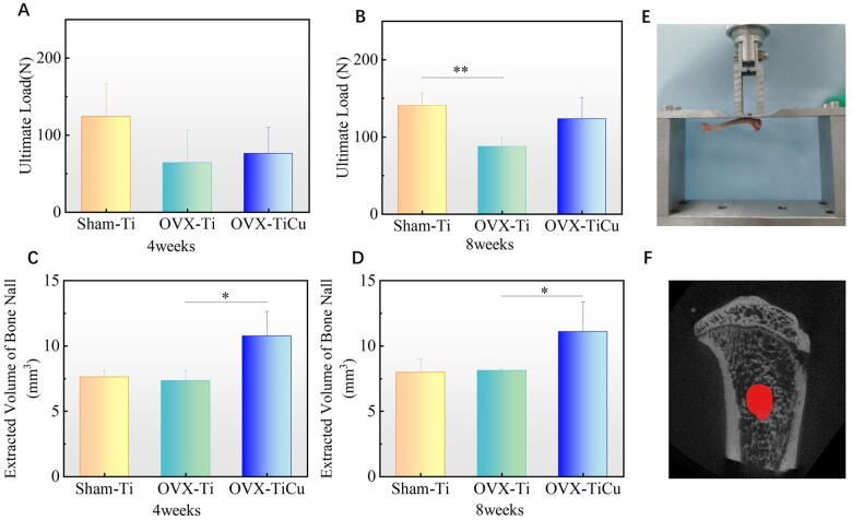
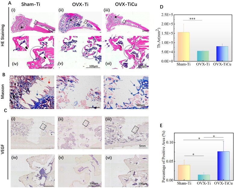
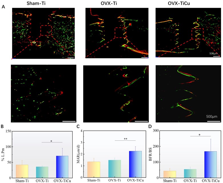
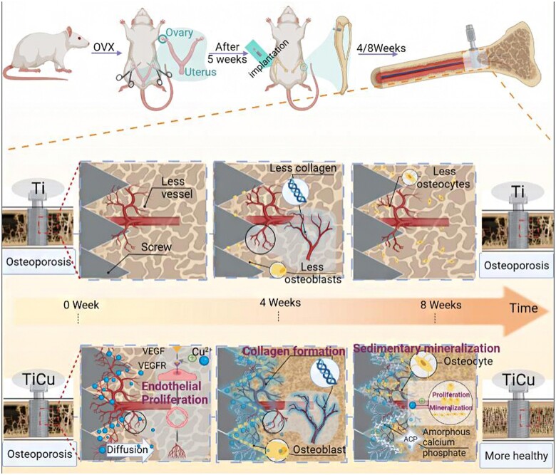
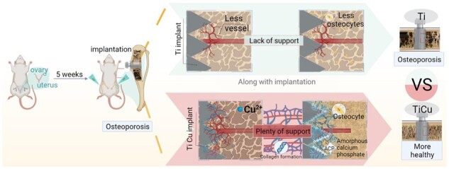
Osteoporosis is a common skeletal disease making patients be prone to the osteoporotic fracture. However, the clinical implants made of titanium and its alloys with a poor osseointegration need a long time for healing and easily to loosening. Thus, a new class of Cu-alloyed titanium (TiCu) alloys with excellent mechanical properties and bio-functionalization has been developed. In this study, the osteoporosis modeled rats were used to study the osteointegration effect and underlying mechanism of TiCu. The results showed that after implantation for 4 weeks, TiCu alloy could promote the reconstruction of vascular network around the implant by up-regulating vascular endothelial growth factor expression. After 8 weeks, it could further promote the proliferation and differentiation of osteoblasts, mineralization and deposition of collagens, and then significantly increasing bone mineral density around the implant. In conclusion, TiCu alloy would enhance the fixation stability, accelerate the osteointegration, and thus reduce the risk of aseptic loosening during the long-term implantation in the osteoporosis environment. This study was the first to report the role and mechanism of a Cu-alloyed metal in promoting osteointegration in osteoporosis environment, which provides a new attractive support for the improvement of future clinical applications of Cu-alloyed antibacterial titanium alloys.
Keywords: copper-alloyed titanium alloy; implant; osseointegration; osteoporosis; vascularization.
© The Author(s) 2022. Published by Oxford University Press.
Figures
References
-
- Arceo-Mendoza RM, Camacho P.. Prediction of fracture risk in patients with osteoporosis: a brief review. Womens Health (Lond) 2015;11:477–82. - PubMed
-
- Ammann P, Rizzoli R.. Bone strength and its determinants. Osteoporos Int 2003;14:S13–8. - PubMed
-
- Li K, Gong H, Xie R, Gu J, Wang S, Lin C, Yin J, Hou X, Zhang Q, Li L, Hao Y.. Clinical efficacy of zoledronic acid combined with percutaneous kyphoplasty in the prevention and treatment of osteoporotic vertebral compression fracture: a systematic review and meta-analysis. Medicine (Baltimore) 2021;100:e25215. - PMC - PubMed
-
- Wang Y, Ma Z, Zheng Y, Liu B, Bao P, Wu X, Yu C, Wen Z, Ma T, Liu J, Liu C, Ma D, Wu H, Li J, Yuan Y, Lu N, Zhao H, Li Y, Yang S, Zhang R, Dai J, Hu M.. Establishment of an osteoporosis model in tree shrews by bilateral ovariectomy and comprehensive evaluation. Exp Ther Med 2019;17:3644–54. - PMC - PubMed
-
- Cu ZY, Meng XY, Feng H, Zhuang SY, Liu ZR, Zhu TJ, Ye KF, Xing Y, Sun C, Zhou F, Tian Y.. Estimation and projection about the standardized prevalence of osteoporosis in mainland China. Arch Osteoporos 2019;15:2. - PubMed
LinkOut - more resources
Full Text Sources
